Copyright
©The Author(s) 2022.
World J Clin Cases. Apr 6, 2022; 10(10): 3156-3163
Published online Apr 6, 2022. doi: 10.12998/wjcc.v10.i10.3156
Published online Apr 6, 2022. doi: 10.12998/wjcc.v10.i10.3156
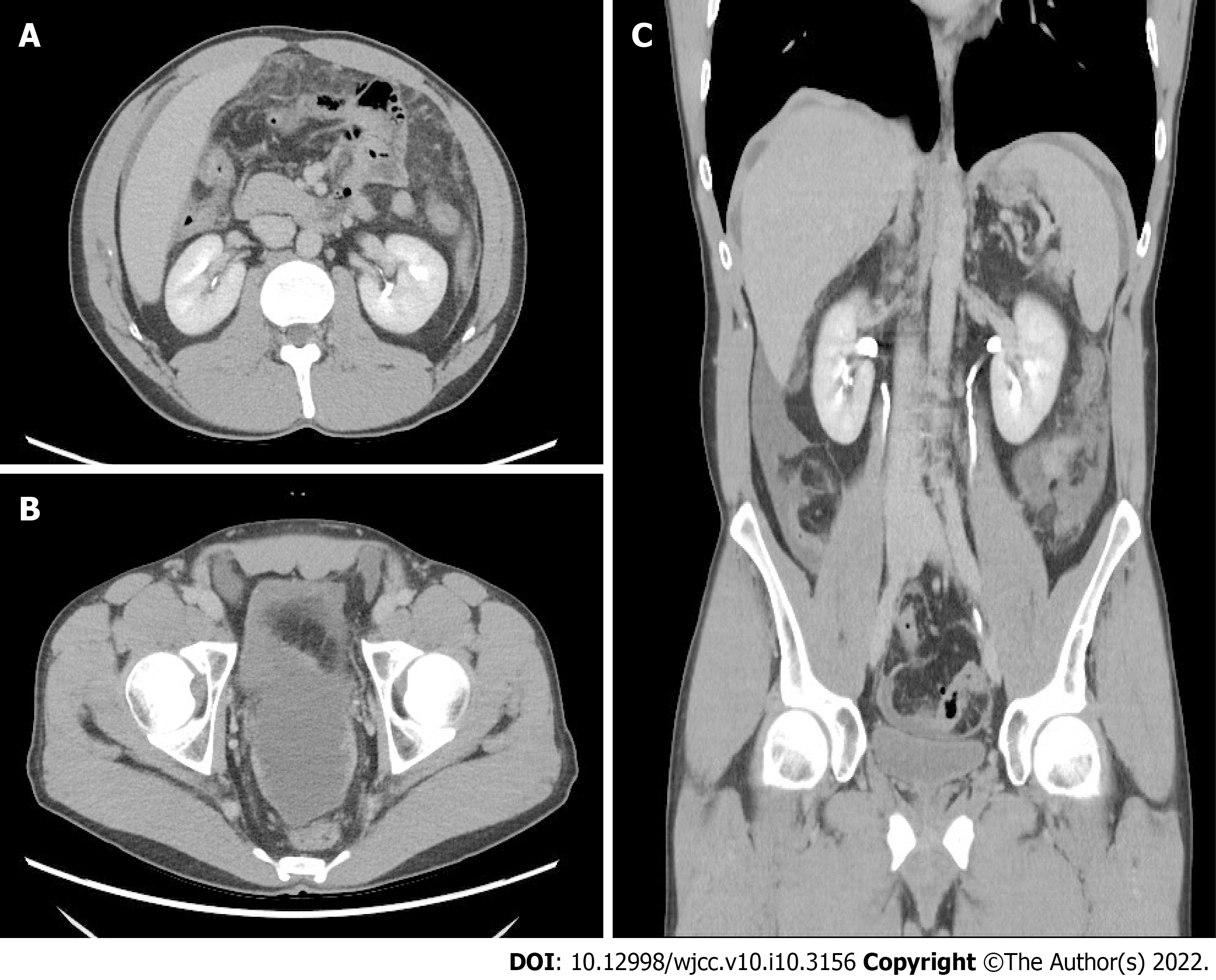
Figure 1 Abdominal contrast-enhanced computed tomography.
A: Axial view, contrast-enhanced computed tomography (CT) showed diffuse fat stranding infiltration within the greater omentum; B: Axial view, thickening of the pelvic peritoneum was enhanced by contrast material; C: Coronal view of the contrast-enhanced CT showed irregular thickening of the perihepatic peritoneum and minimal ascites over the bilateral subphrenic spaces, paracolic gutter, and pelvic cavity.
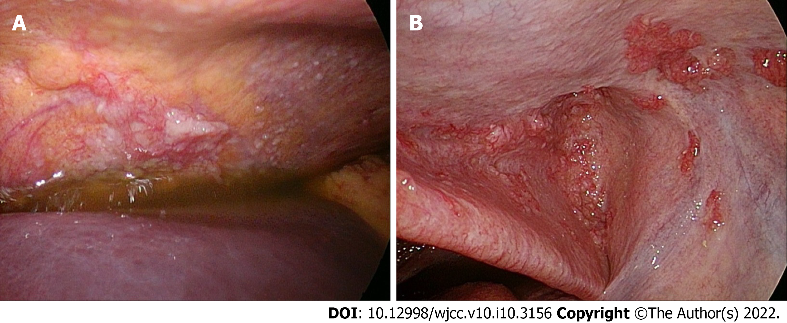
Figure 2 Laparoscopy.
A: Multiple whitish nodules were observed over the entire abdominal cavity with some yellow ascites; B: Grossly red nodular tissue was located on the peritoneum.
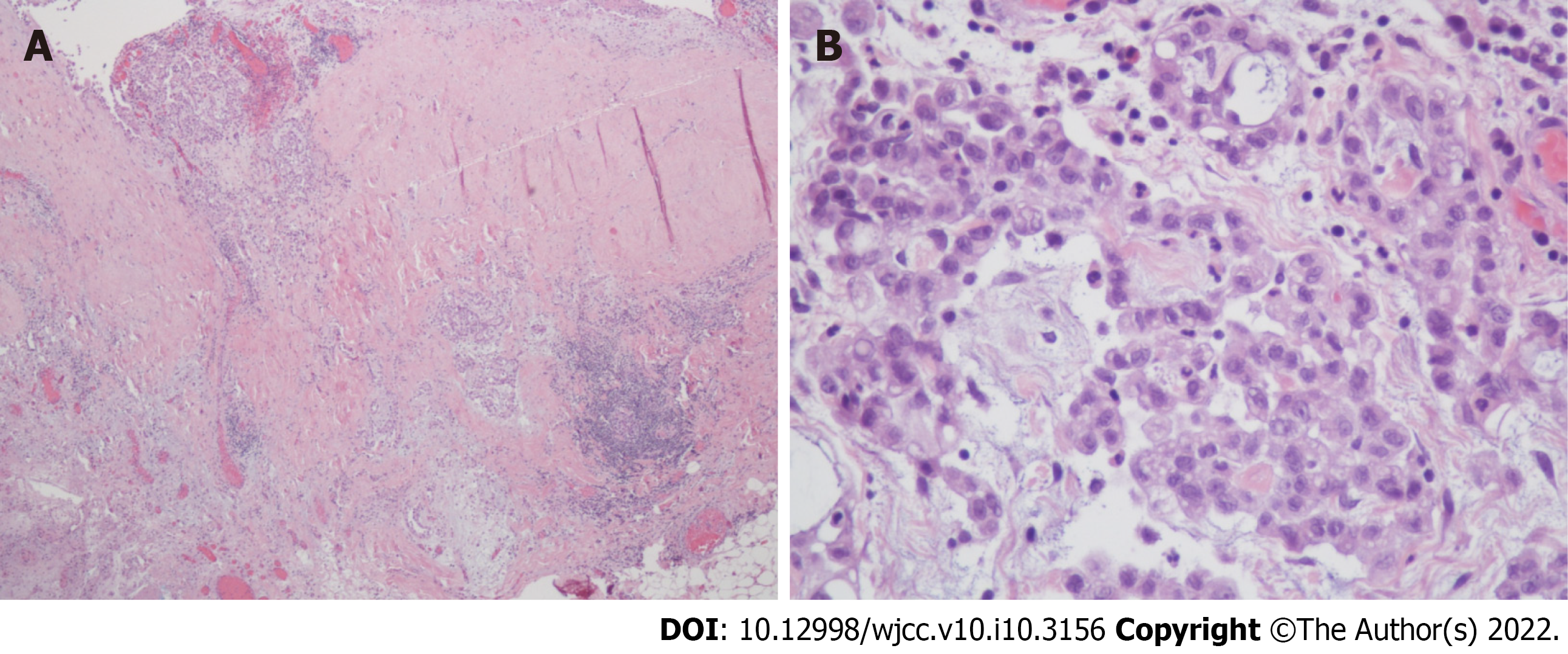
Figure 3 Pathological findings of the lesion on the peritoneum.
A: The peritoneum revealed mesothelioma invasion into the stroma and adipose tissue composed of a tubular or single-cell arrangement of epithelioid cells (x 40; HE stain); B: The tumor cells had large, oval-round nuclei, conspicuous nucleoli, and moderate eosinophilic cytoplasm (x 400; HE stain).
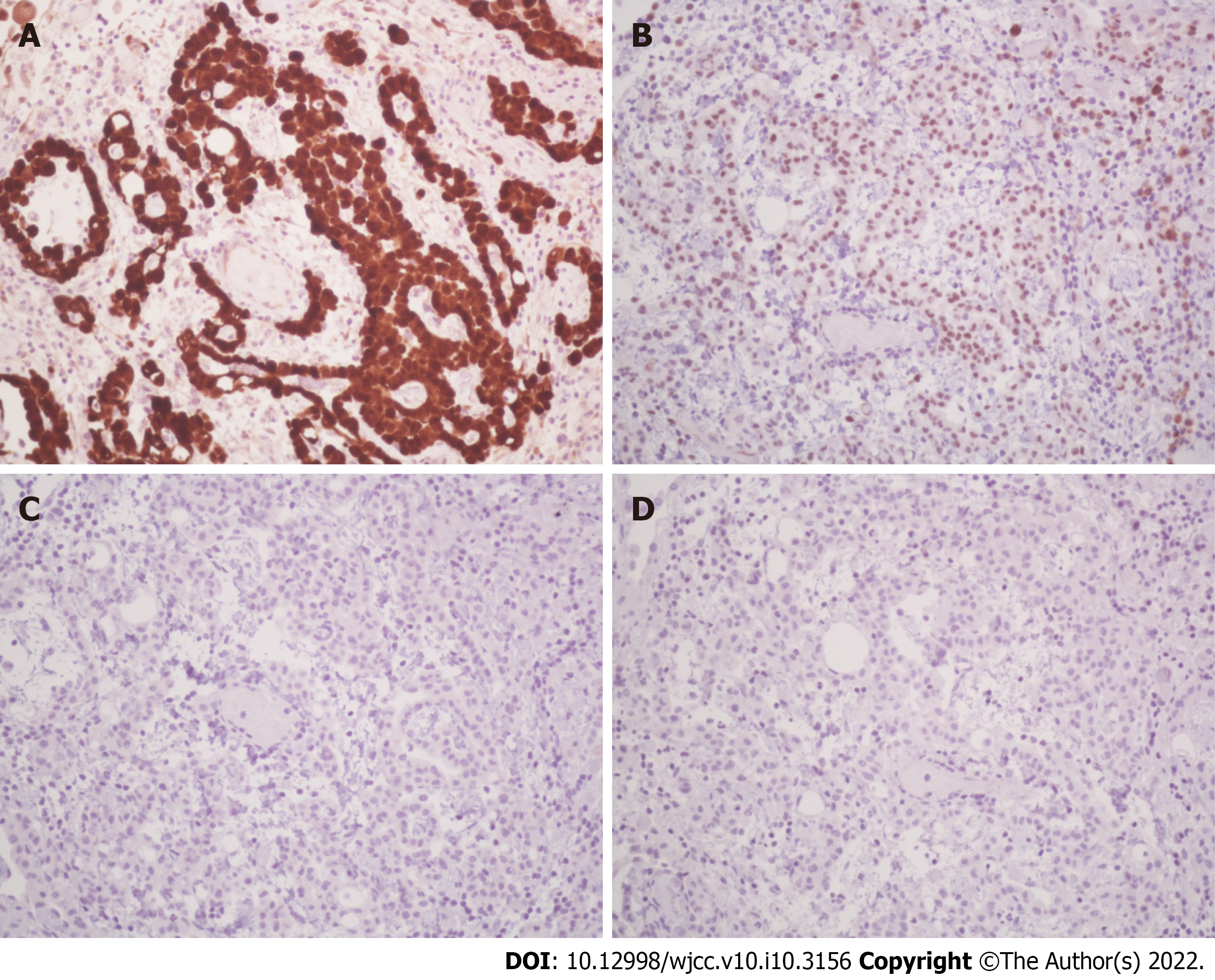
Figure 4 The immunohistochemical staining results.
A: Immunohistochemically, the tumor cells were positive for calretinin (nuclear and cytoplasmic staining, x 200); B: The tumor cells were focally positive for WT-1 (x 200); C: TTF-1 immunostaining was negative for tumor cells (x 200); D: CDX-2 immunostaining was negative for tumor cells (x200).
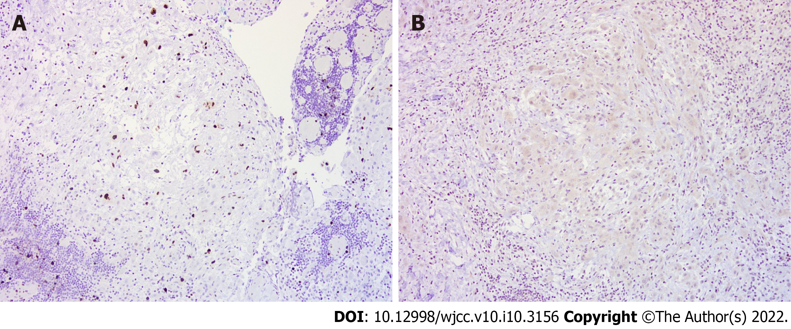
Figure 5 Additional staining results.
A: Ki-67 proliferative index was < 10%(x 100); B: Napsin A immunostaining was negative for tumor cell (x 100).
- Citation: Lin LC, Kuan WY, Shiu BH, Wang YT, Chao WR, Wang CC. Primary malignant peritoneal mesothelioma mimicking tuberculous peritonitis: A case report. World J Clin Cases 2022; 10(10): 3156-3163
- URL: https://www.wjgnet.com/2307-8960/full/v10/i10/3156.htm
- DOI: https://dx.doi.org/10.12998/wjcc.v10.i10.3156


