Copyright
©The Author(s) 2022.
World J Clin Cases. Apr 6, 2022; 10(10): 3014-3026
Published online Apr 6, 2022. doi: 10.12998/wjcc.v10.i10.3014
Published online Apr 6, 2022. doi: 10.12998/wjcc.v10.i10.3014
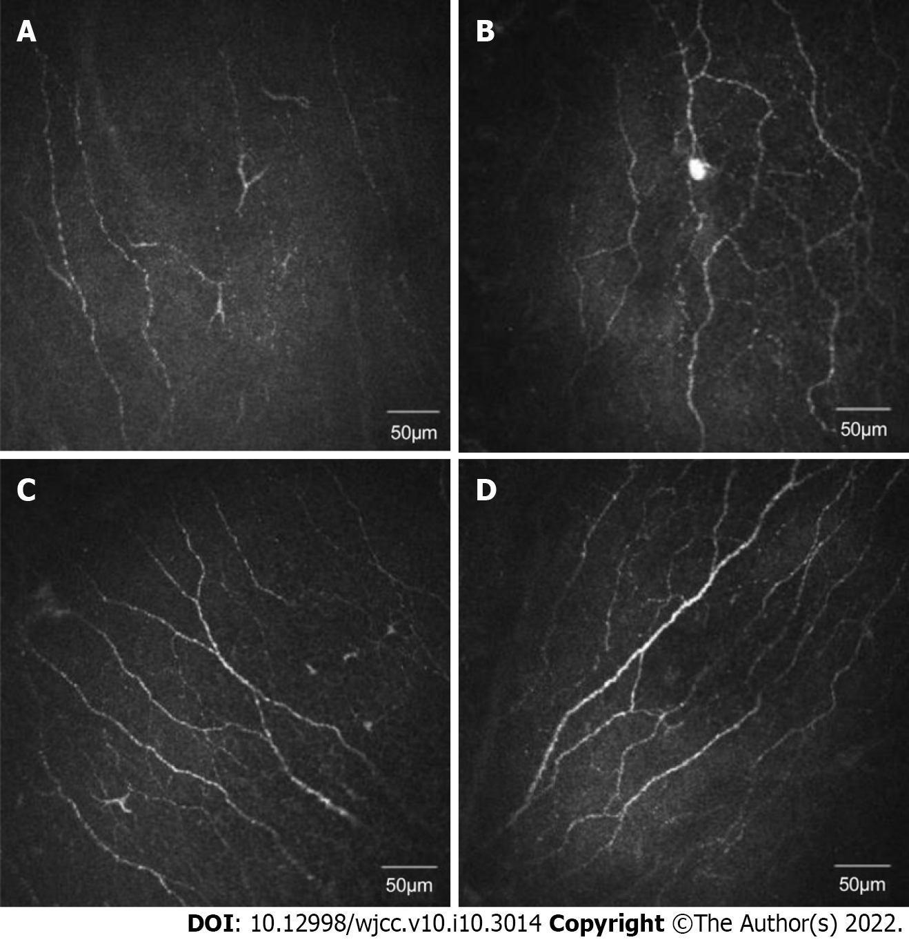
Figure 1 In vivo confocal microscopy of corneal sub-basal nerves of four representative patients.
A: A dry eye patient with diabetes; B: A dry eye patient without diabetes; C: A diabetes patient without dry eye; D: A control subject. The size of each image is 400 μm × 400 μm.
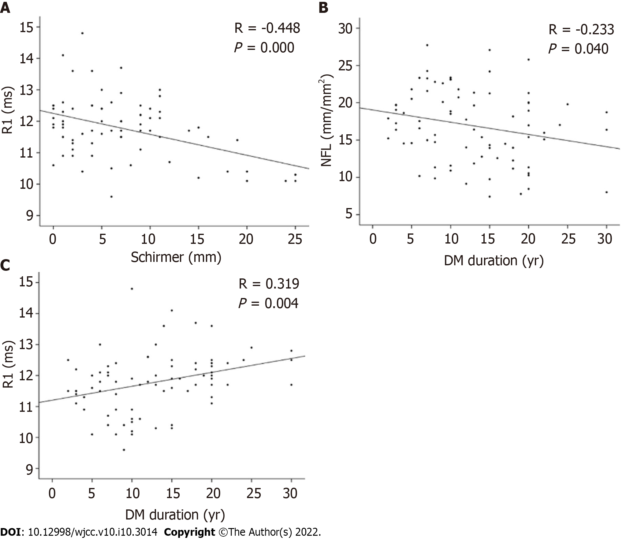
Figure 2 Nerve fiber length was also associated with diabetes duration.
A: Correlation between R1 Latency and the Schirmer I score; B: Correlation between nerve fiber length and diabetes duration time; C: Correlation between R1 Latency and diabetes duration time. NFL: Nerve fiber length. DM: Diabetes mellitus.
- Citation: Fang W, Lin ZX, Yang HQ, Zhao L, Liu DC, Pan ZQ. Changes in corneal nerve morphology and function in patients with dry eyes having type 2 diabetes. World J Clin Cases 2022; 10(10): 3014-3026
- URL: https://www.wjgnet.com/2307-8960/full/v10/i10/3014.htm
- DOI: https://dx.doi.org/10.12998/wjcc.v10.i10.3014


