Copyright
©The Author(s) 2022.
World J Clin Cases. Jan 7, 2022; 10(1): 381-387
Published online Jan 7, 2022. doi: 10.12998/wjcc.v10.i1.381
Published online Jan 7, 2022. doi: 10.12998/wjcc.v10.i1.381
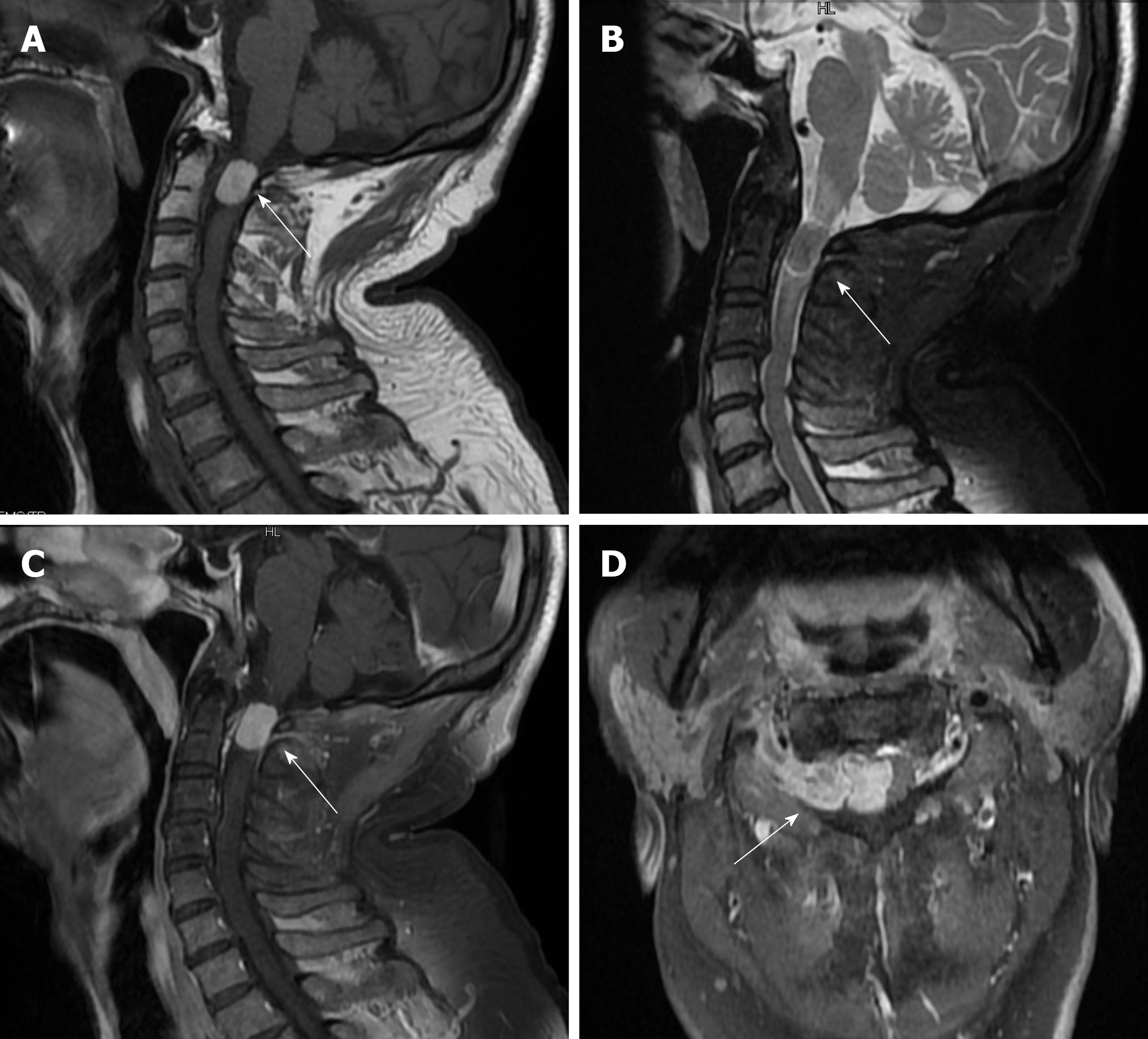
Figure 1 A: T1-weighted showed high-intensity; B: T2-weighted was equal-signal; C: Contrast Enhancement showed clear boundary, and the spinal cord is significantly compressed and displaced to the left side; D: White arrow the tumor grew out of spinal canal through intervertebral foramen.
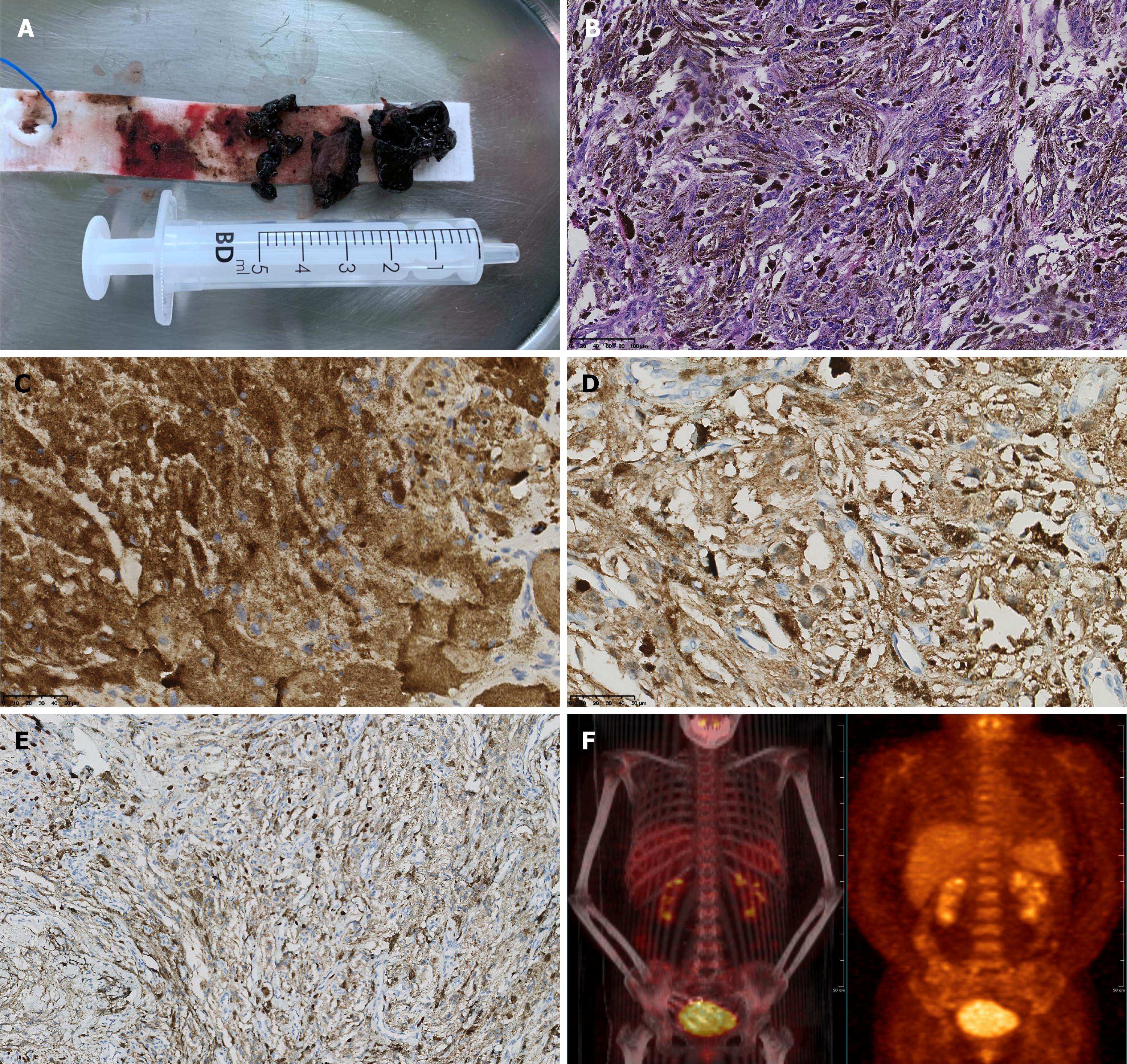
Figure 2 A: A black mass (tumor) was tough and solid, lack of blood supply; B: Hematoxylin-eosin stain (HE, original magnification ×200); C: HMB45(+); D: S100(+); E: Ki67 (approximately 20%); F: Positron emission tomography/ computerized tomography scan showed negative.
- Citation: Shi YF, Chen YQ, Chen HF, Hu X. An atypical primary malignant melanoma arising from the cervical nerve root: A case report and review of literture. World J Clin Cases 2022; 10(1): 381-387
- URL: https://www.wjgnet.com/2307-8960/full/v10/i1/381.htm
- DOI: https://dx.doi.org/10.12998/wjcc.v10.i1.381


