Copyright
©The Author(s) 2022.
World J Clin Cases. Jan 7, 2022; 10(1): 331-337
Published online Jan 7, 2022. doi: 10.12998/wjcc.v10.i1.331
Published online Jan 7, 2022. doi: 10.12998/wjcc.v10.i1.331
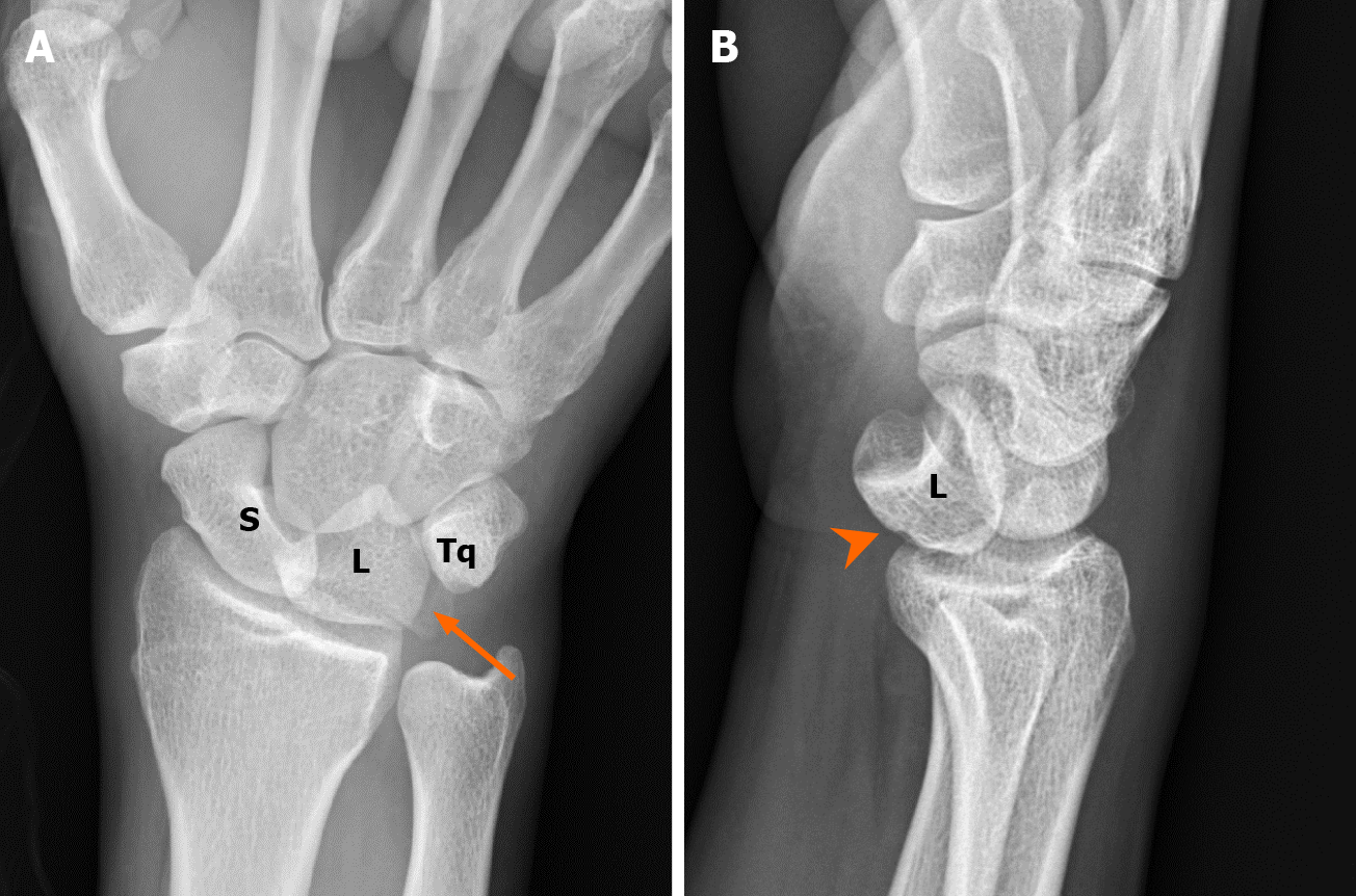
Figure 1 A 37-year-old man with multiple traumas.
Radiological examinations revealed acute lunate dislocation with triquetral fracture. A: Plain radiograph in the emergency department showed triangular appearance of the lunate (piece of pie) with triquetral fracture. Note the avulsion of the triquetrum (arrow) due to failure of the lunotriquetral ligaments; B: Lateral view of the radiograph showed volar dislocation of the lunate bone (arrowhead). S: Scaphoid, L: Lunate, Tq: Triquetrum.
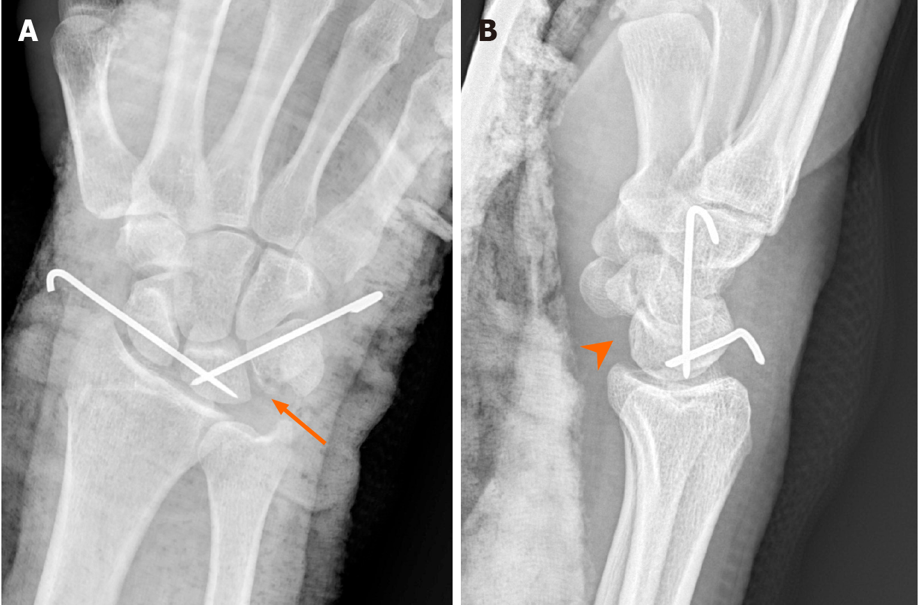
Figure 2 Post closed reduction and fixation with two Kirschner wires.
A: The postoperative plain radiograph showed appropriate reduction of the lunate with the smooth alignment of the great arc. Note the avulsed triquetrum (arrow) reduced by ligamentotaxis of the lunotriquetral ligament; B: Lateral view of the radiograph showed appropriate reduction with neutral scapholunate angle (arrowhead).
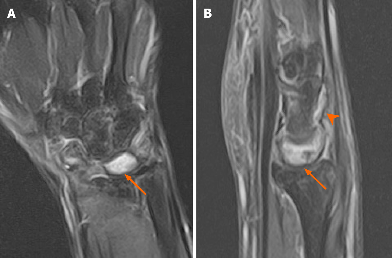
Figure 3 Two months postoperative magnetic resonance imaging showed residual bone edema of the lunate.
A: Magnetic resonance imaging (MRI) in T2W coronal Fat saturation (FS) showed residual bone marrow edema in scaphoid, triquetrum and particularly, the lunate (arrow). This finding might be attributed to the post-traumatic or postoperative change; B: MRI in T2W sagittal FS showed lunate bone marrow edema and effusion in the intercarpal joints (arrowhead).
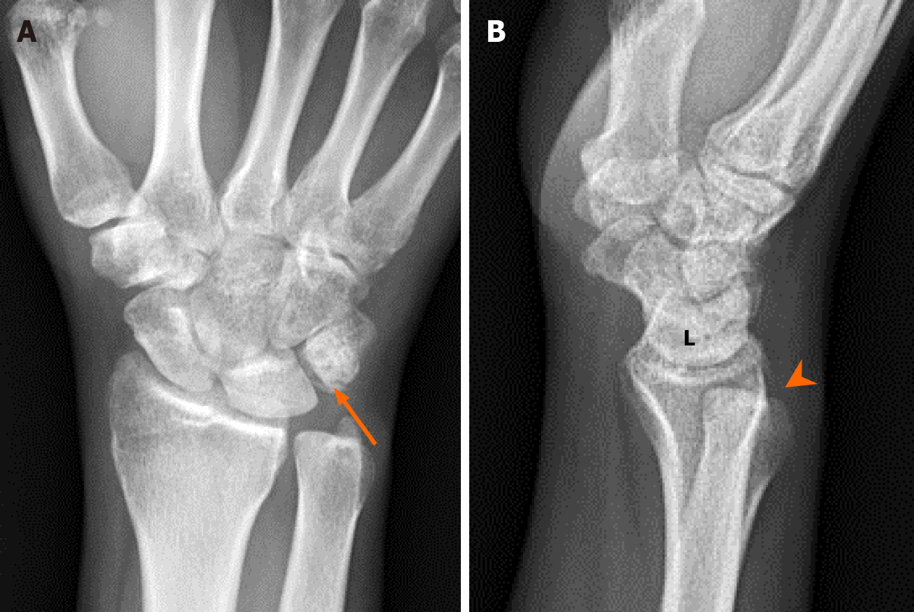
Figure 4 Five months postoperative plain radiograph showed appropriate lunate alignment.
A: The radiograph showed solid union of the avulsed triquetrum (arrow) with no associated widening of the scapholunate and lunotriquetral joint space, and no necrotic changes in the lunate; B: Lateral view of the wrist joint showed a neutral scapholunate ligament with no obvious volar or dorsal intercalated segment instability. However, residual distal radioulnar joint subluxation was observed during follow-up (arrowhead).
Figure 5 Wrist motion range six months postoperatively.
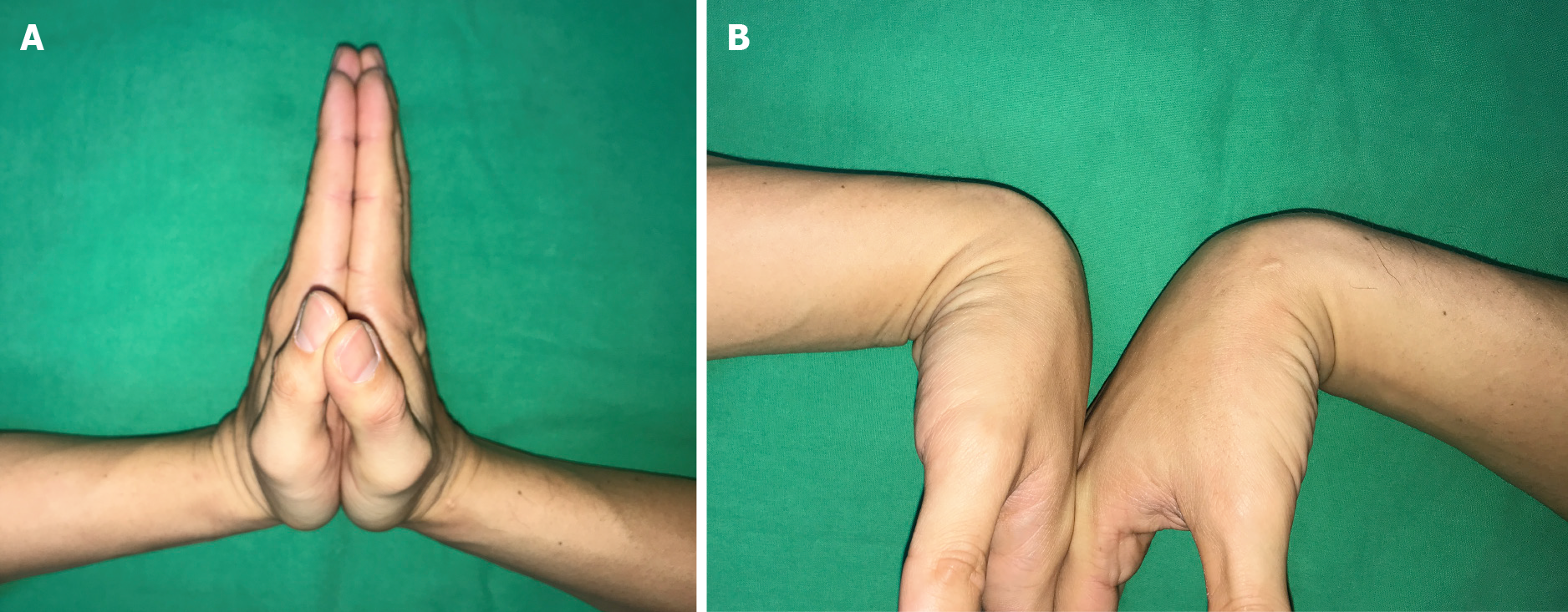
A: With 90° extension; B: 75° flexion.
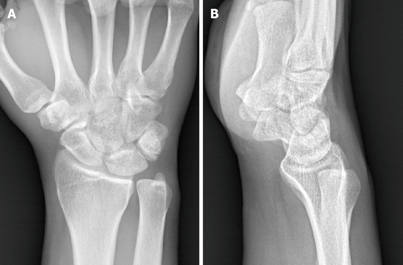
Figure 6 One year postoperative plain radiograph showed appropriate lunate alignment.
A: Anteroposterior view of the wrist joint; B: Lateral view of the wrist joint. There was no evidence of avascular necrosis of the lunate, scapholunate instability, or carpal joint arthritis.
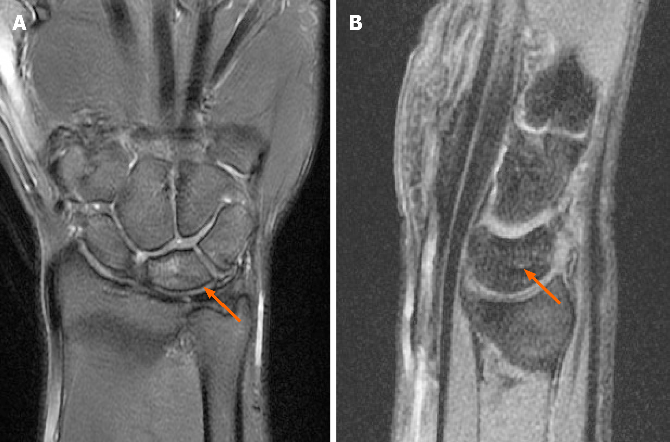
Figure 7 Magnetic resonance imaging follow-up 18 mo postoperatively.
A: Coronal section; B: Sagittal section. These findings showed almost completely resorption of bone edema of the lunate (arrow) and did not progress into osteonecrosis.
- Citation: Li LY, Lin CJ, Ko CY. Lunate dislocation with avulsed triquetral fracture: A case report. World J Clin Cases 2022; 10(1): 331-337
- URL: https://www.wjgnet.com/2307-8960/full/v10/i1/331.htm
- DOI: https://dx.doi.org/10.12998/wjcc.v10.i1.331


