Copyright
©The Author(s) 2022.
World J Clin Cases. Jan 7, 2022; 10(1): 309-315
Published online Jan 7, 2022. doi: 10.12998/wjcc.v10.i1.309
Published online Jan 7, 2022. doi: 10.12998/wjcc.v10.i1.309
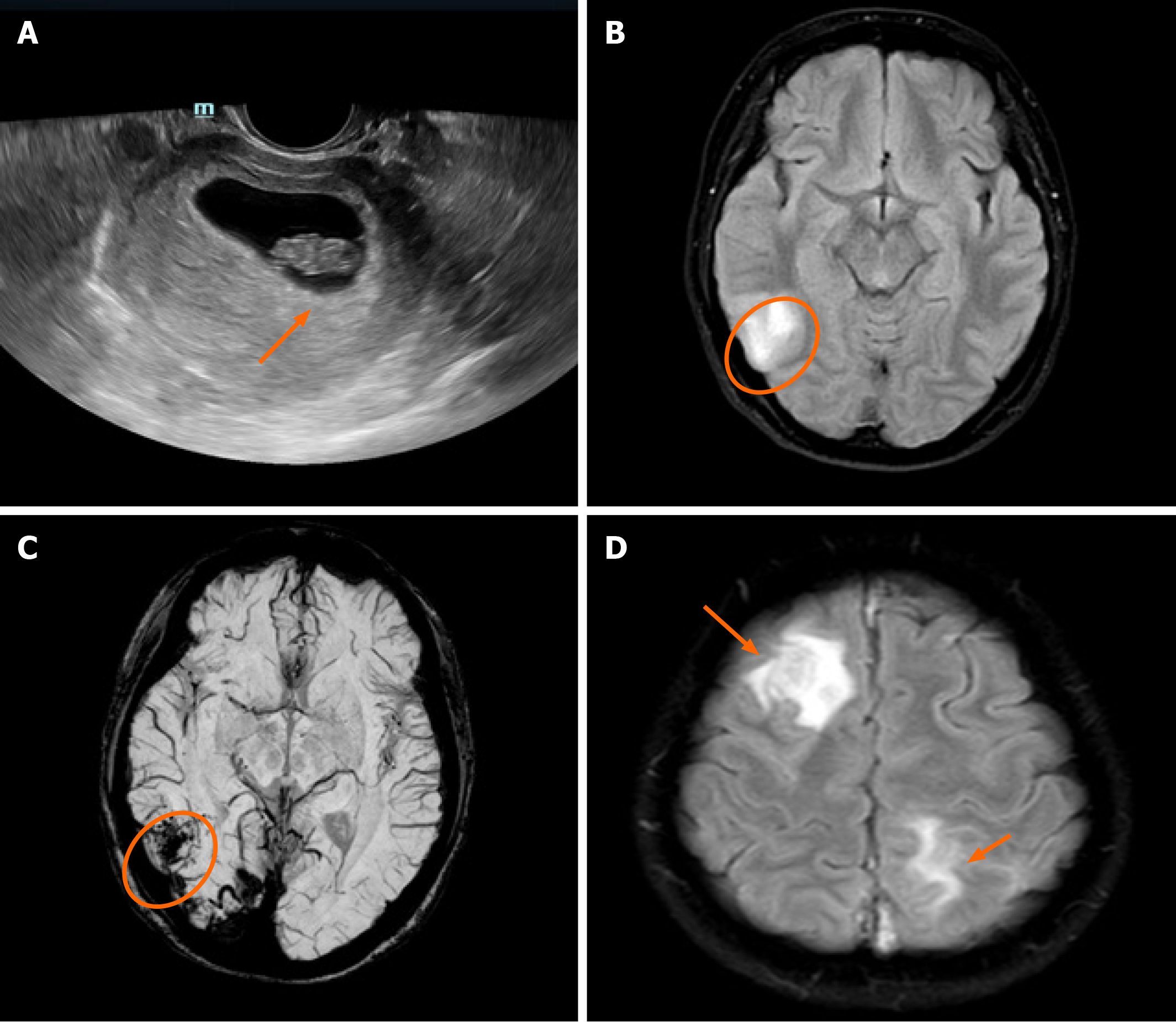
Figure 1 Ultrasound and magnetic resonance imaging.
A: Ultrasound shows a fetus at 8 wk of gestation (orange arrow); B: Pre-thrombectomy-diffusion weighted imaging shows restriction in the right temporal lobe (orange circle); C: Pre-thrombectomy susceptibility-weighted imaging shows a large area of low signal region in the right temporal lobe; D: Post-thrombectomy magnetic resonance imaging reveals new lesions in the right frontal lobe and left parietal lobe (orange arrows).
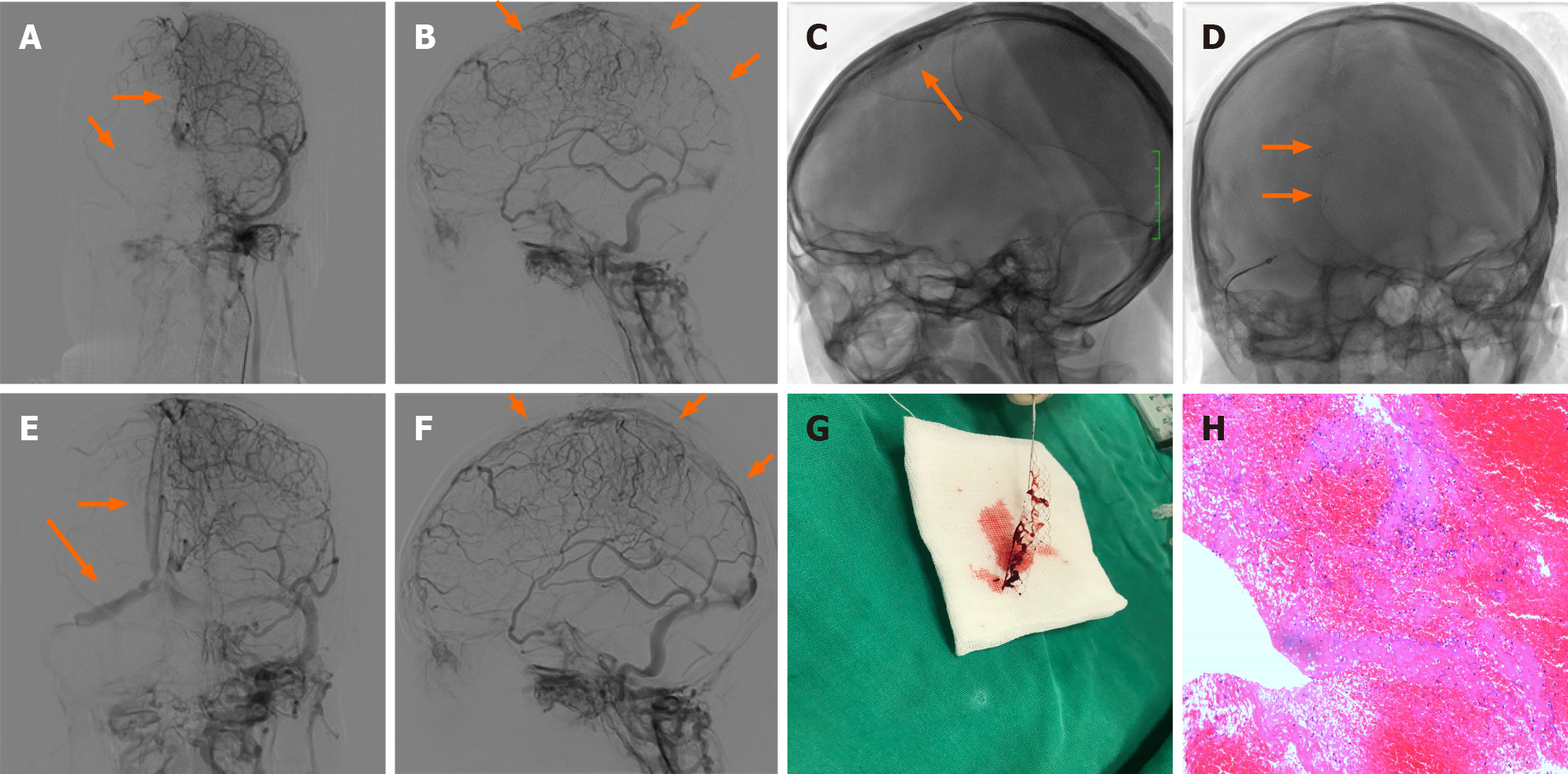
Figure 2 Operating process and outcome.
A and B: Prethrombectomy angiogram confirms occlusion of superior longitudinal sinus, as well as right transverse sinus, and sinus sigmoideus (orange arrows); C and D: Mechanical thrombectomy was performed using two Solitaire stents (orange arrows); E and F: Post-thrombectomy angiogram shows resolution of flow in the superior longitudinal sinus, right transverse sinus and sinus sigmoideus (orange arrows); G: Gross appearance of the partial removed cerebral embolus; H: Microscopic appearance of the cerebral embolus specimen consistent with mixed thrombus.
- Citation: Zhou B, Huang SS, Huang C, Liu SY. Cerebral venous sinus thrombosis in pregnancy: A case report . World J Clin Cases 2022; 10(1): 309-315
- URL: https://www.wjgnet.com/2307-8960/full/v10/i1/309.htm
- DOI: https://dx.doi.org/10.12998/wjcc.v10.i1.309


