Copyright
©The Author(s) 2022.
World J Clin Cases. Jan 7, 2022; 10(1): 289-295
Published online Jan 7, 2022. doi: 10.12998/wjcc.v10.i1.289
Published online Jan 7, 2022. doi: 10.12998/wjcc.v10.i1.289
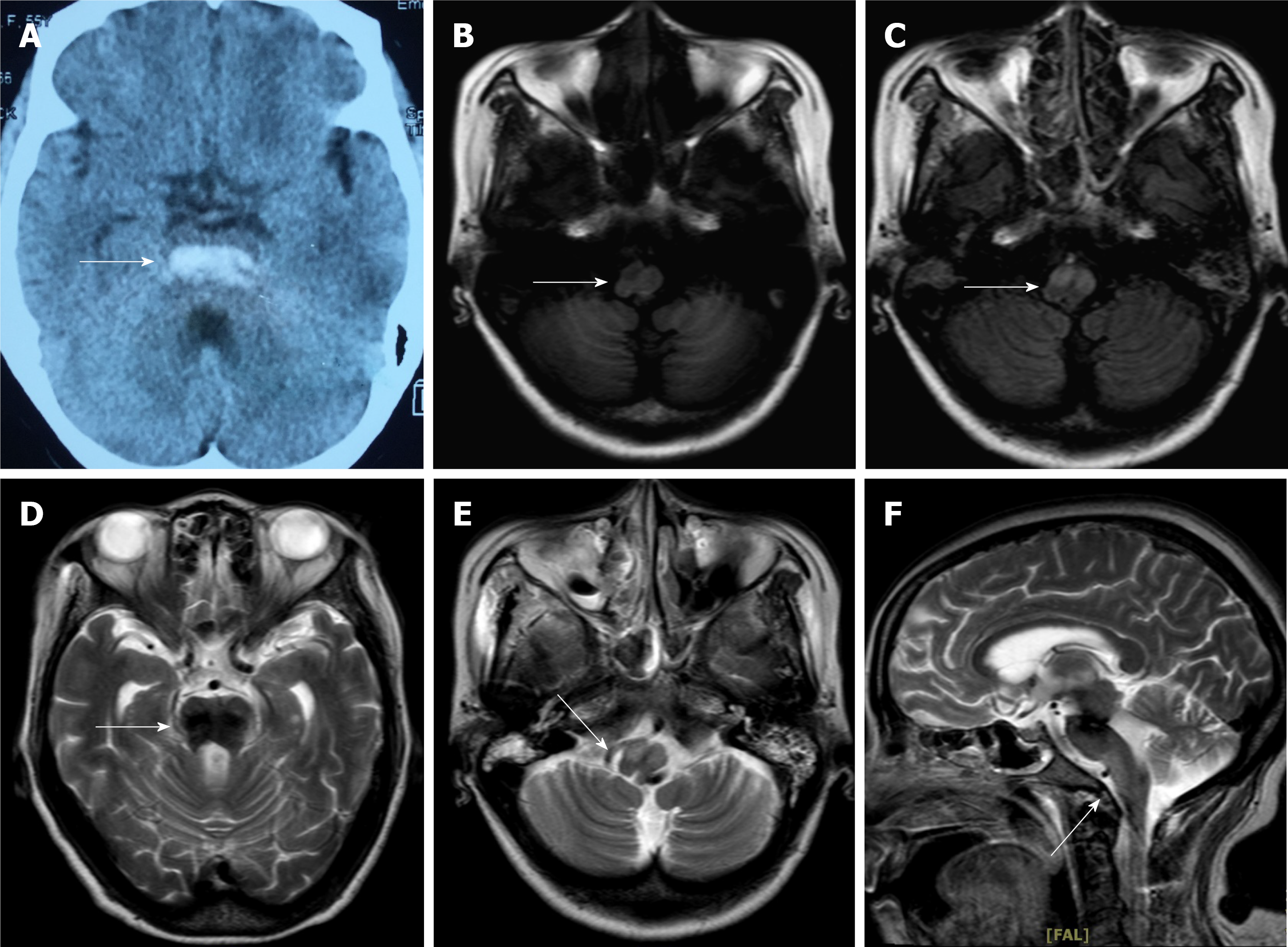
Figure 1 The results of brain computed tomography 3 months ago and magnetic resonance imaging (MRI) after admission.
A: The results of the brain computed tomography demonstrate an acute bilateral pontine haemorrhage; B: The results of T2-weighted axial MRI show the residual blood region after 3 months; C: The results of T1-weighted axial MRI through the medulla demonstrate expansion of the bilateral inferior olivary nucleus; D-F: The white arrow region shows hyperintense signal in the T2-weighted and fluid-attenuated inversion-recovery (FLAIR) sequence, and it displays no enhancement or restricted diffusion on others. (D: Axial T2-weighted image; E: Axial FLAIR image; F: Sagittal T2-weighted image).
- Citation: Zheng B, Wang J, Huang XQ, Chen Z, Gu GF, Luo XJ. Bilateral Hypertrophic Olivary Degeneration after Pontine Hemorrhage: A Case Report. World J Clin Cases 2022; 10(1): 289-295
- URL: https://www.wjgnet.com/2307-8960/full/v10/i1/289.htm
- DOI: https://dx.doi.org/10.12998/wjcc.v10.i1.289


