Copyright
©The Author(s) 2022.
World J Clin Cases. Jan 7, 2022; 10(1): 268-274
Published online Jan 7, 2022. doi: 10.12998/wjcc.v10.i1.268
Published online Jan 7, 2022. doi: 10.12998/wjcc.v10.i1.268
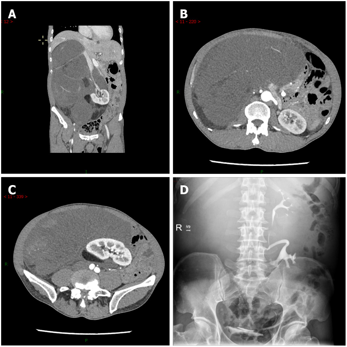
Figure 1 Imaging findings before treatment.
A and C: Computed tomography angiography showed a massive lipoma-like mass extending from the sub-hepatic space to the pelvic cavity, with multiple organs dislocated; B and D: Intravenous pyelography with radiocontrast agent confirmed the displacement of the right kidney to the left lower quadrant and its excretion function was good.
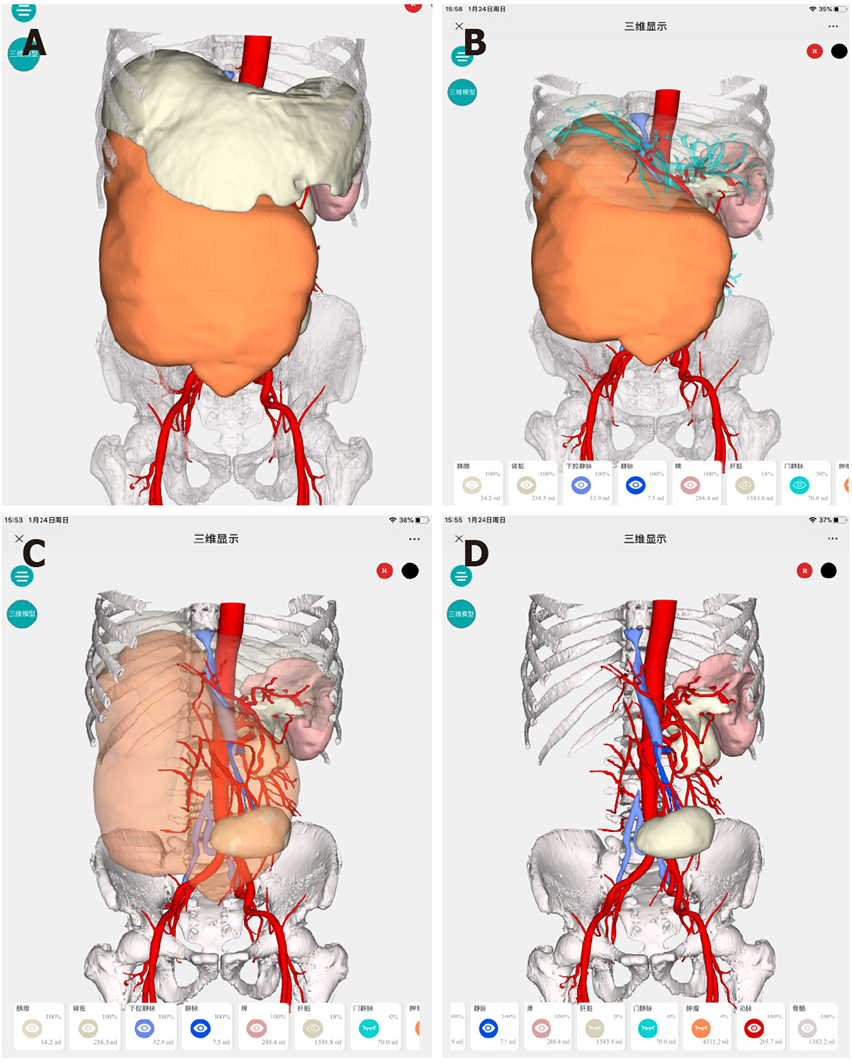
Figure 2 Hyper-accuracy three-dimensional reconstruction.
A: Three-dimensional surface-rendered organs and tumor; B: Semitransparentizing liver revealed the relationship between the tumor and liver; C and D: Semitransparentizing or hiding tumor revealed the detail variations regarding anatomical structures. More details: http://www.cas.hisense.com:10052/?id=VnVmamZNSzhCbl8zODQ1&type=newCode.
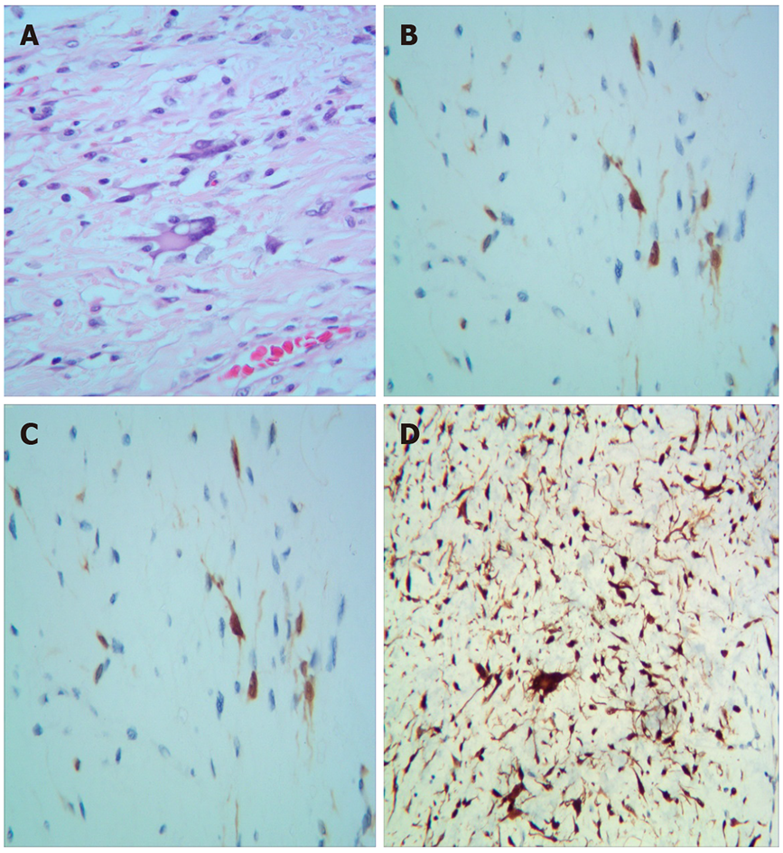
Figure 3 Pathological examination results.
A: Pathological examination revealed well-differentiated liposarcoma. Macroscopically, the mass appeared oval but was separated into irregular lobulations; B-E: The epithelial component showed a low grade of dedifferentiation. Immunohistochemically, the tumor was partly positive for MDM2, S100, CK34, and CKD4, with a low grade of dedifferentiation (Ki-67: 20%).
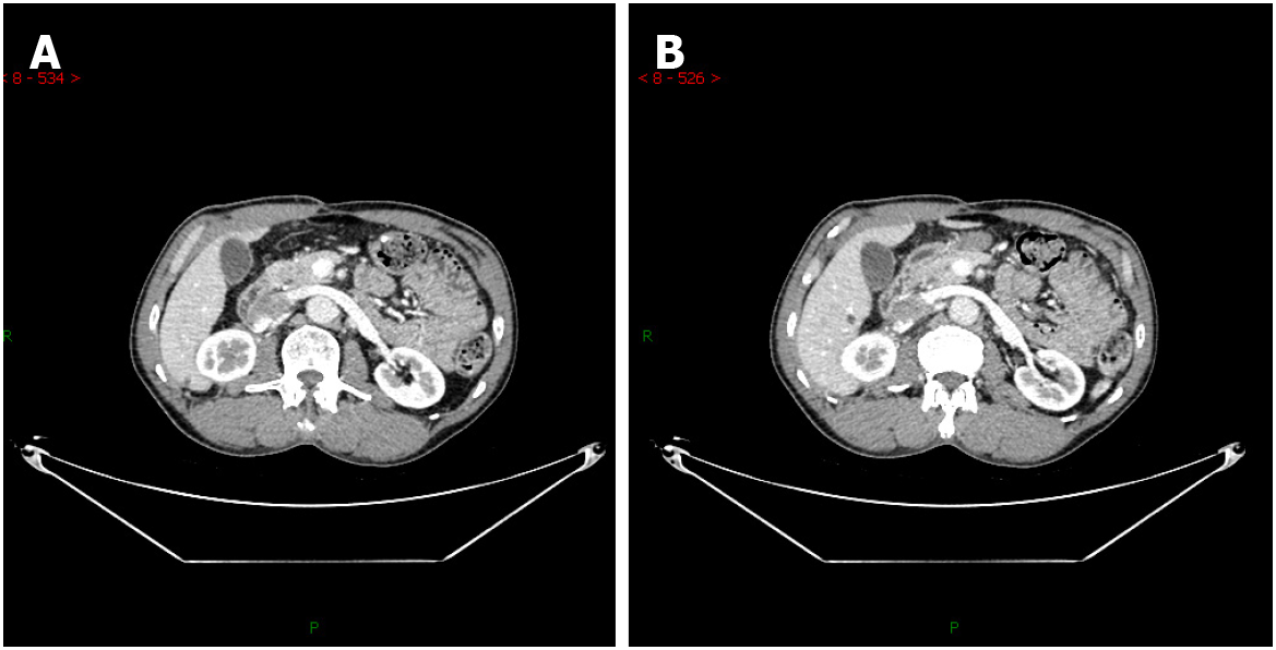
Figure 4 Contrast enhanced computed tomography images.
A: Contrast enhanced computed tomography (CT) 3 mo after surgery; B: Contrast enhanced CT 16 mo after surgery.
- Citation: Ye MS, Wu HK, Qin XZ, Luo F, Li Z. Hyper-accuracy three-dimensional reconstruction as a tool for better planning of retroperitoneal liposarcoma resection: A case report. World J Clin Cases 2022; 10(1): 268-274
- URL: https://www.wjgnet.com/2307-8960/full/v10/i1/268.htm
- DOI: https://dx.doi.org/10.12998/wjcc.v10.i1.268


