Copyright
©The Author(s) 2022.
World J Clin Cases. Jan 7, 2022; 10(1): 260-267
Published online Jan 7, 2022. doi: 10.12998/wjcc.v10.i1.260
Published online Jan 7, 2022. doi: 10.12998/wjcc.v10.i1.260
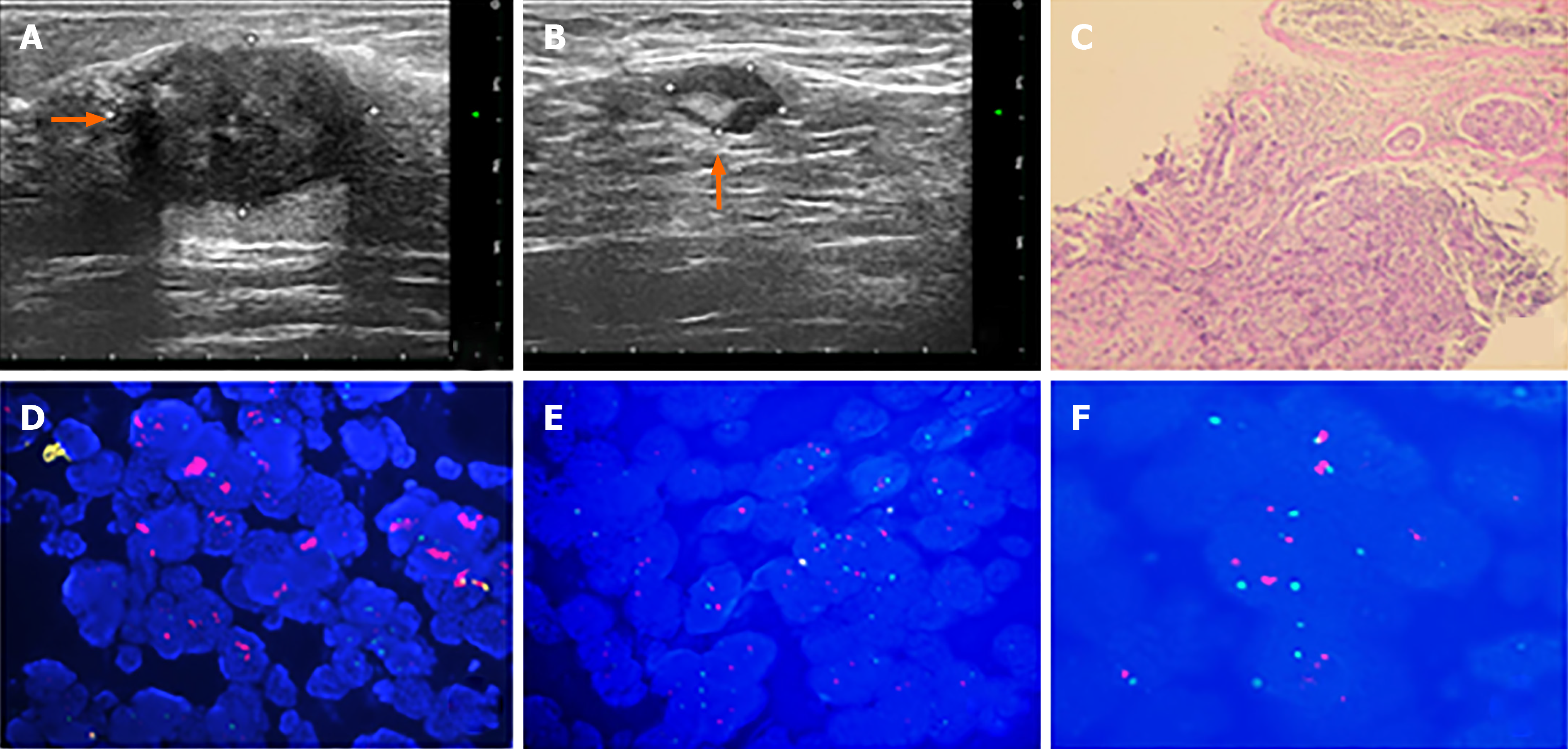
Figure 1 Malignant tumor in the right breast when initially diagnosed.
A: Ultrasound showing the mass measuring 2.73 cm × 2.13 cm × 2.57 cm in the 10-o'clock position of the right breast; B: Ultrasound showing the largest right axillary lymph node measuring 1.2 cm × 0.9 cm; C: Hematoxylin-eosin staining indicates an invasive ductal carcinoma (×100); D: Positive control of fluorescence in situ hybridization (FISH); E: Negative control of FISH; F: FISH result of the biopsy specimen.
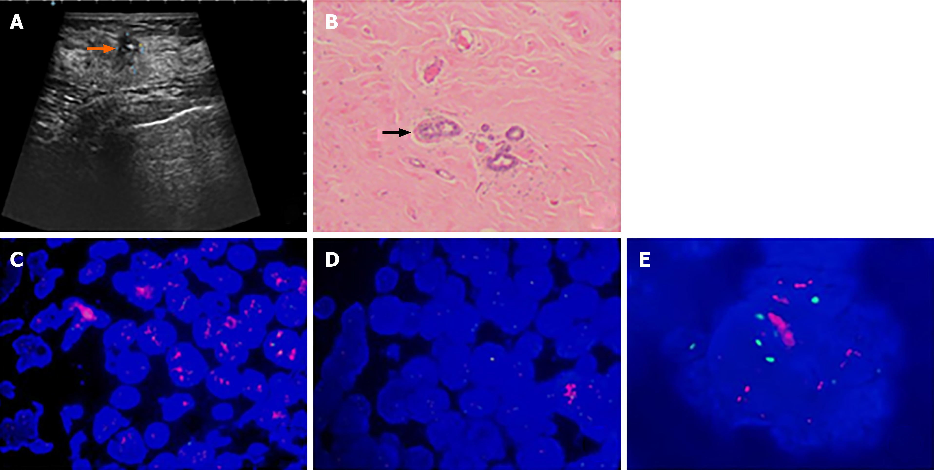
Figure 2 Ultrasound result after neoadjuvant chemotherapy and histopathological and fluorescence in situ hybridization findings of the resected tumor.
A: Ultrasound demonstrating the tumor measuring 0.8 cm × 0.7 cm after neoadjuvant chemotherapy (arrow); B: Histopathological finding showing the tumor cells tubular arrangement and the development of invasion (H&E staining, × 50) (arrow); C: Positive control of fluorescence in situ hybridization (FISH); D: Negative control of FISH; E: FISH result of the surgically resected tumor.
- Citation: Wang L, Jiang Q, He MY, Shen P. HER2 changes to positive after neoadjuvant chemotherapy in breast cancer: A case report and literature review . World J Clin Cases 2022; 10(1): 260-267
- URL: https://www.wjgnet.com/2307-8960/full/v10/i1/260.htm
- DOI: https://dx.doi.org/10.12998/wjcc.v10.i1.260


