Copyright
©The Author(s) 2022.
World J Clin Cases. Jan 7, 2022; 10(1): 143-154
Published online Jan 7, 2022. doi: 10.12998/wjcc.v10.i1.143
Published online Jan 7, 2022. doi: 10.12998/wjcc.v10.i1.143
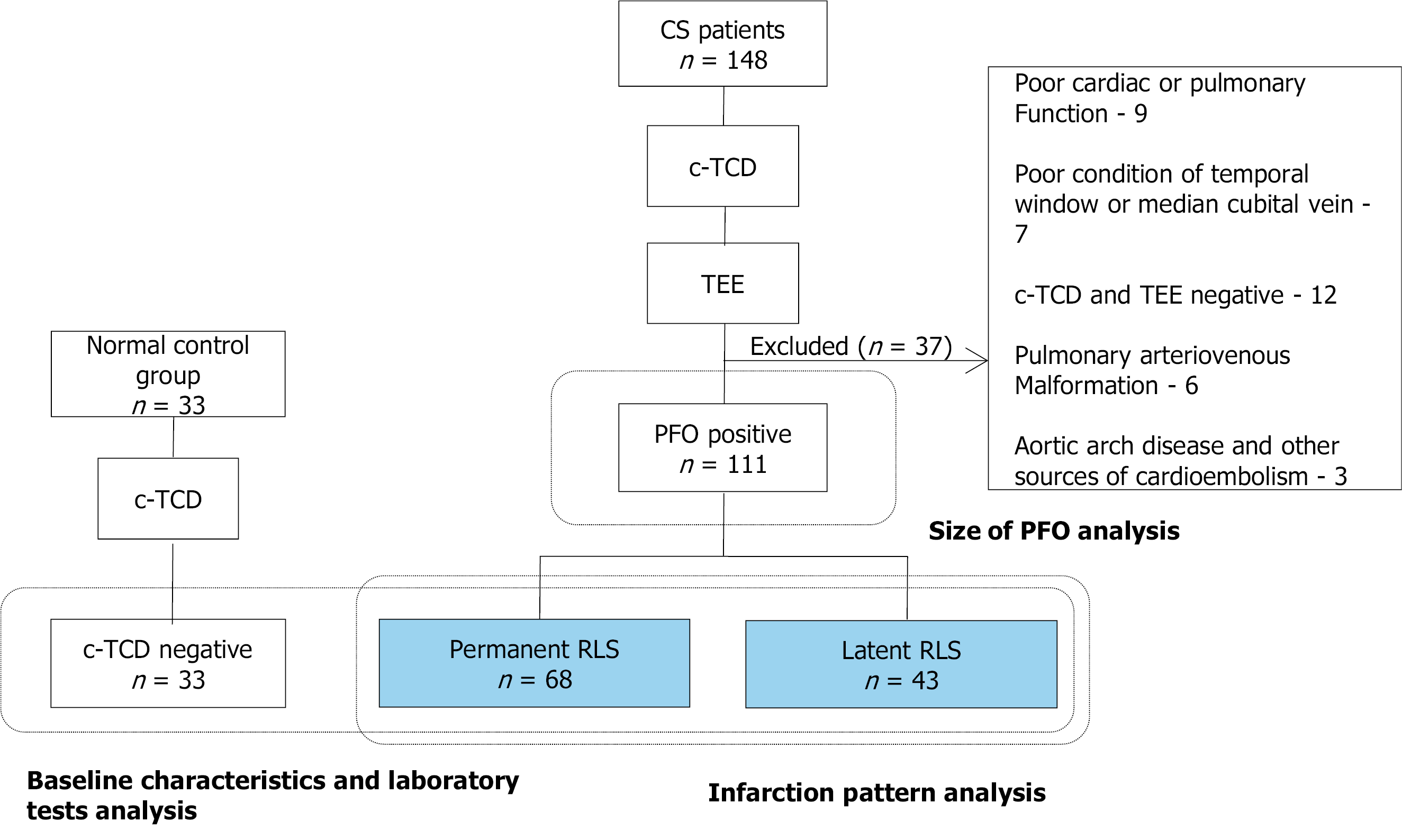
Figure 1 Flowchart of patient selection.
CS: Cryptogenic stroke; c-TCD: Contrast-enhanced transcranial Doppler; PFO: Patent foramen ovale; RLS: Right-to-left shunt; TEE: Transesophageal echocardiography.
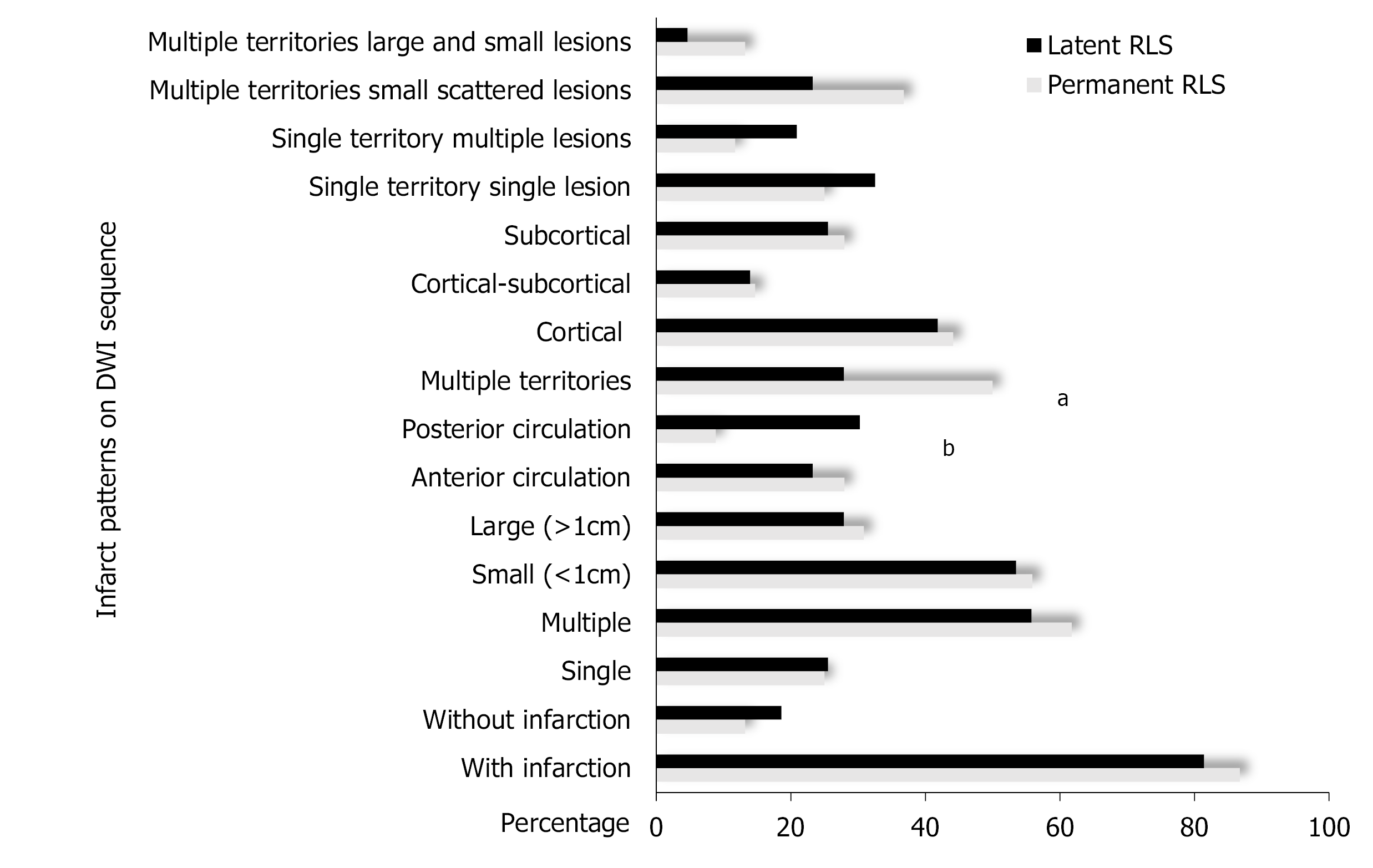
Figure 2 Diffusion-weighted imaging lesions of patent foramen ovale-related cryptogenic stroke patients with permanent and latent right-to-left shunt.
The lesions in permanent right-to-left shunt (RLS) were more likely to be distributed in multiple territories, while the lesions of latent RLS were more likely to be distributed in the posterior circulation. aP < 0.05; bP < 0.01. DWI: Diffusion-weighted imaging.
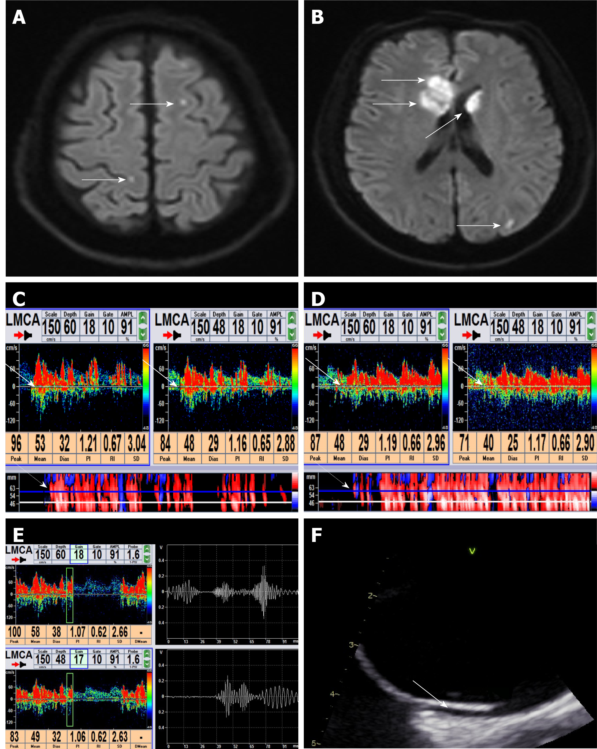
Figure 3 Diffusion-weighted imaging, contrast-enhanced transcranial Doppler and transesophageal echocardiography images of a patient.
A-B: Diffusion-weighted imaging of brain magnetic resonance imaging showed multiple infarcts on the left superior frontal gyrus and right precentral gyrus (A, white solid arrows), left occipital gyrus and the head of bilateral caudate and corpus callosum (B, white solid arrows); C-E: Contrast-enhanced transcranial Doppler images; C: The left middle cerebral artery was monitored in single-channel dual-depth mode. Large microbubble signals (Grade III) were detected at a resting state. Microbubbles were detected as high-intensity transient signals within the Doppler flow spectrum at 60 mm and 48 mm monitoring depths (white solid arrows). Microbubble signals were also detected on M-mode (white dotted arrow); D: Curtain microbubble signals (Grade IV) were detected during conventional Valsalva maneuver, and the white solid arrows show microbubbles as high-intensity transient signals within the doppler flow spectrum at 60 mm and 48 mm monitoring depth. Microbubble signals were also detected on M-mode (white dotted arrow); E: Microbubble signals in green sampling boxes on the left half showed a spike-like appearance on the sonogram on the right half; F: Transesophageal echocardiography showed the patent foramen ovale was a potential fissure between the primary septum and secondary septum, presenting an imbricate structure (white solid arrow).
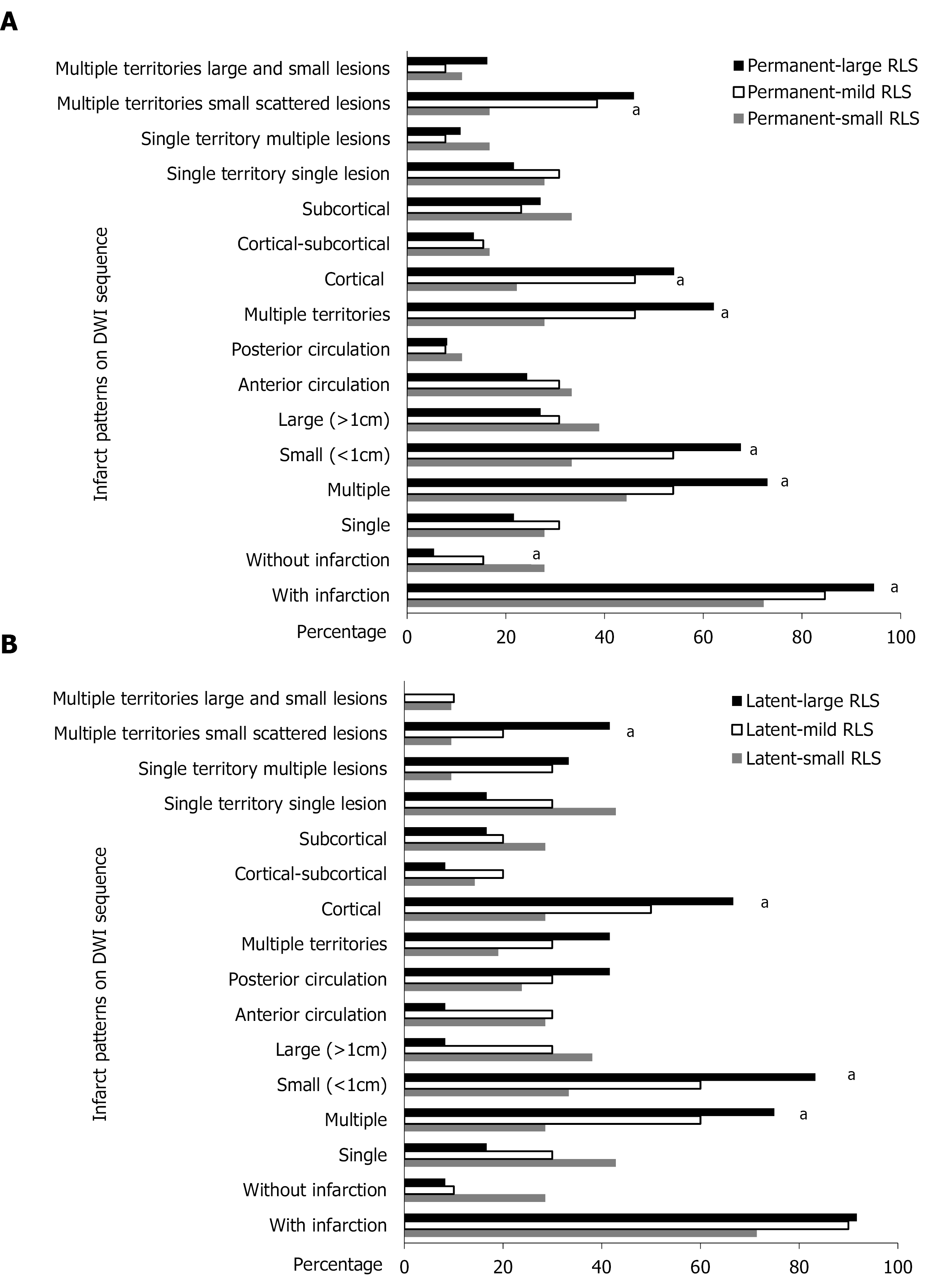
Figure 4 Diffusion-weighted imaging lesions of different grades of shunt in permanent and latent right-to-left shunt groups.
A: Infarction pattern of permanent-small, permanent-mild and permanent-large right-to-left shunt (RLS). Multiple, small and cortical lesions were more likely to be involved as the grade of shunt increased in the permanent RLS group; B: Infarction pattern of latent-small, latent-mild and latent-large RLS. Multiple, small and cortical lesions were more likely to be involved as the grade of shunt increased in the latent RLS group. aP < 0.05.
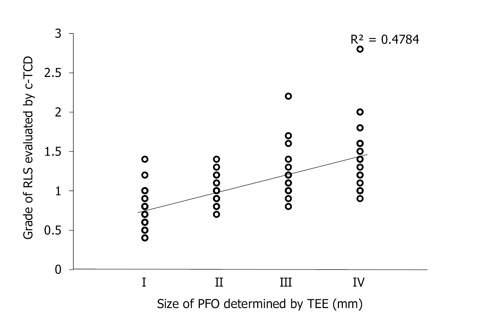
Figure 5 Correlation between the grade of right-to-left shunt on contrast-enhanced transcranial Doppler and the size of patent foramen ovale on transesophageal echocardiography of patent foramen ovale-related cryptogenic stroke patients.
r = 0.758, P < 0.001. c-TCD: Contrast-enhanced transcranial Doppler; PFO: Patent foramen ovale; RLS: Right-to-left shunt; TEE: Transesophageal echocardiography.
- Citation: Xiao L, Yan YH, Ding YF, Liu M, Kong LJ, Hu CH, Hui PJ. Evaluation of right-to-left shunt on contrast-enhanced transcranial Doppler in patent foramen ovale-related cryptogenic stroke: Research based on imaging. World J Clin Cases 2022; 10(1): 143-154
- URL: https://www.wjgnet.com/2307-8960/full/v10/i1/143.htm
- DOI: https://dx.doi.org/10.12998/wjcc.v10.i1.143


