Copyright
©The Author(s) 2022.
World J Clin Cases. Jan 7, 2022; 10(1): 1-11
Published online Jan 7, 2022. doi: 10.12998/wjcc.v10.i1.1
Published online Jan 7, 2022. doi: 10.12998/wjcc.v10.i1.1
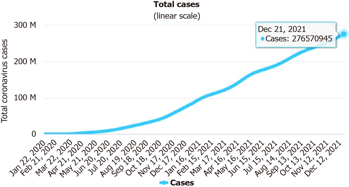
Figure 1 Linear trend of number of severe acute respiratory syndrome coronavirus 2 infected cases (January 22, 2020 to December 21, 2021).
Source: https://www.worldometers.info/coronavirus/#countries.
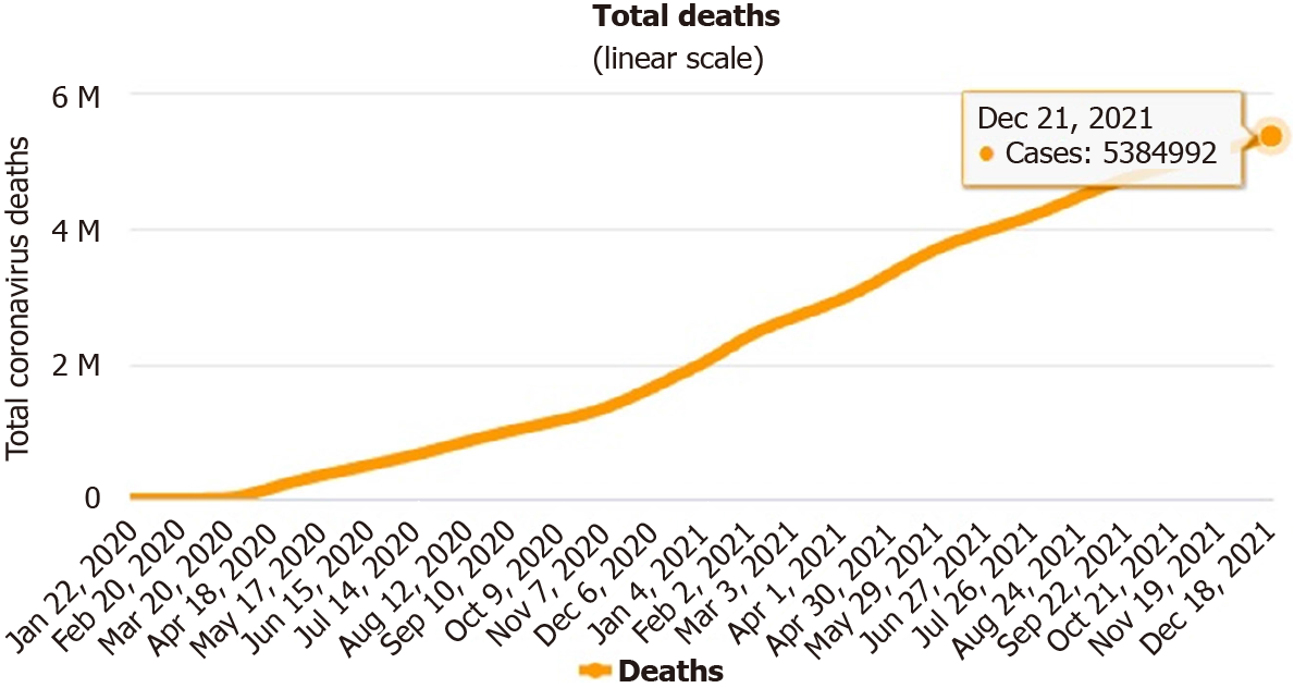
Figure 2 Trend of number of people who died from severe acute respiratory syndrome coronavirus 2 (January 22, 2020 to December 21, 2021).
Source: https://www.worldometers.info/coronavirus/#countries.
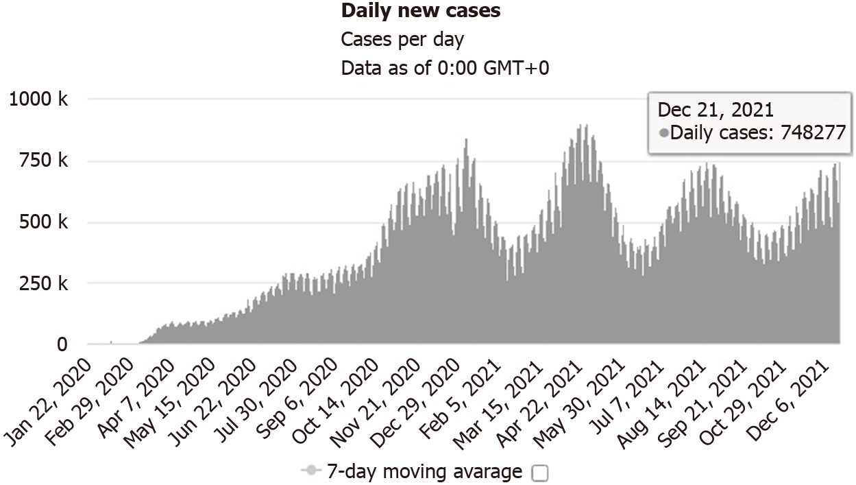
Figure 3 Daily new confirmed severe acute respiratory syndrome coronavirus 2 infected cases (January 22, 2020 to December 21, 2021).
Source: https://www.worldometers.info/coronavirus/#countries.
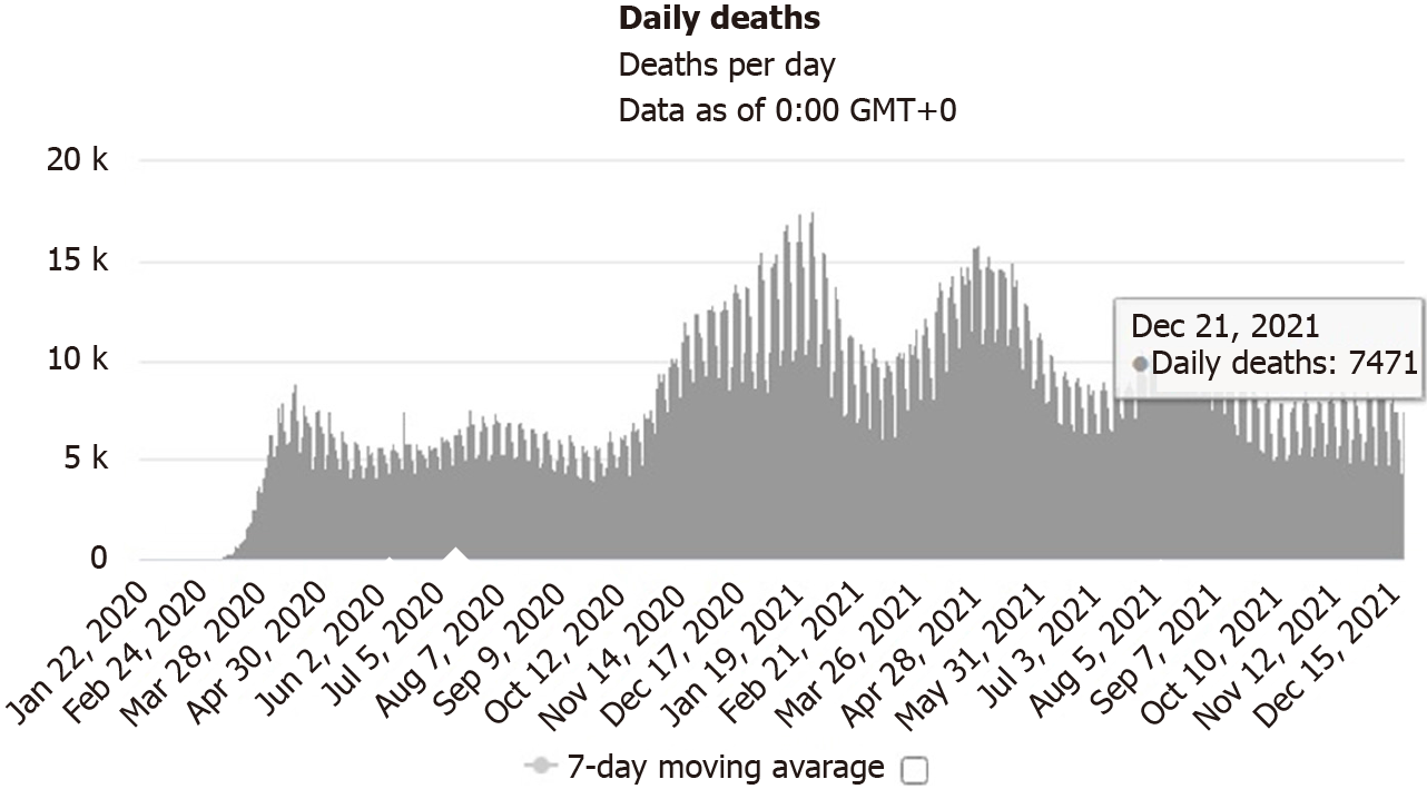
Figure 4 Daily new deaths due to coronavirus disease 2019 (January 22, 2020 to December 21, 2021).
Source: https://www.worldometers.info/coronavirus/#countries.
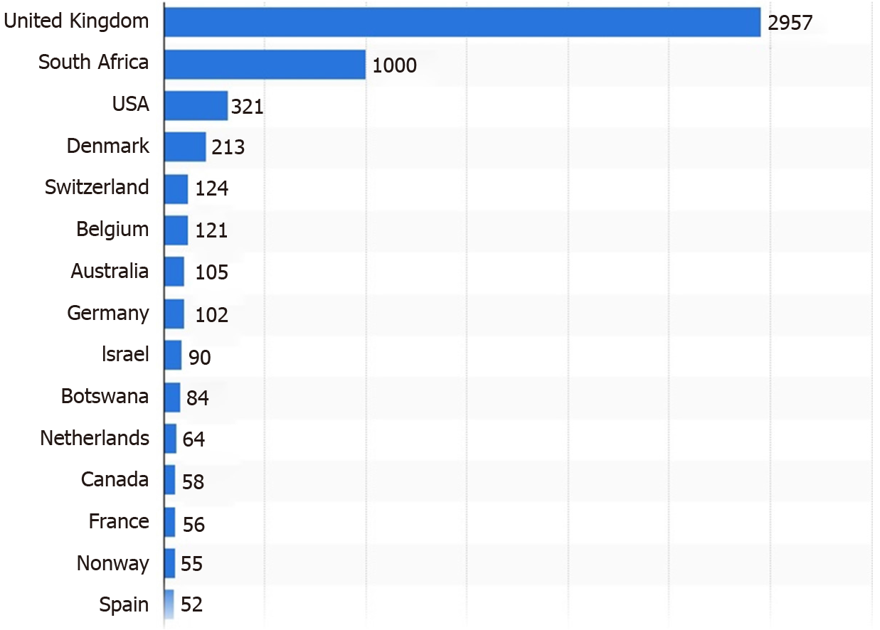
Figure 5 Number of severe acute respiratory syndrome coronavirus 2 Omicron variant cases worldwide as of December 16, 2021.
Source: https://www.statista.com/statistics/1279100/number-omicron-variant-worldwide-by-country/.
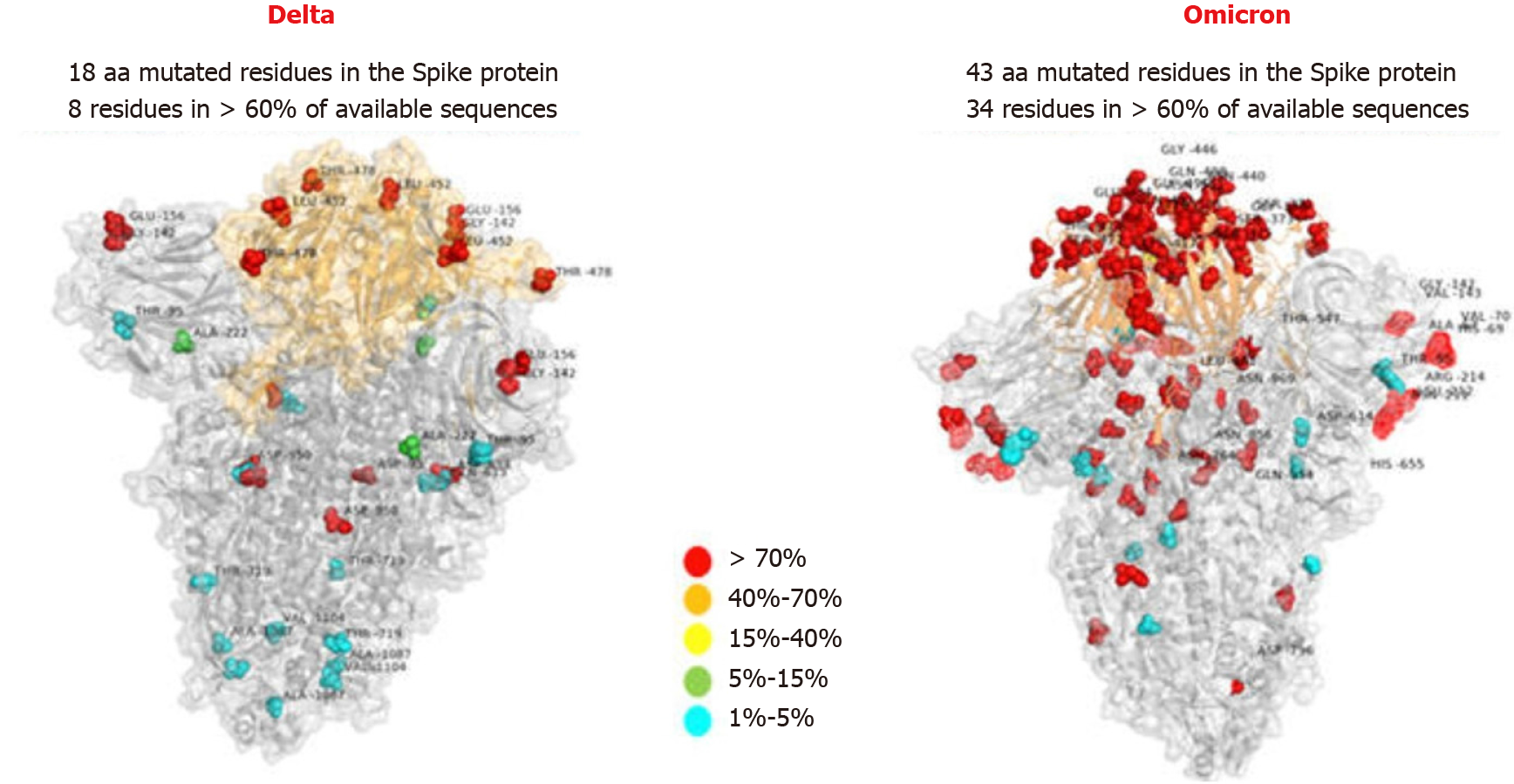
Figure 6 Comparison of structure of spike protein between the Omicron and the Delta of severe acute respiratory syndrome coronavirus 2 issued by Italy Bambino Gesù Children Hospital showing the active site in orange and the residues colored against different mutational rates.
Source: https://new.qq.com/rain/a/20211214A087SY00?refer=wx_hot.
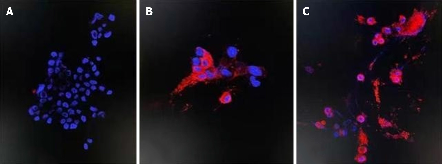
Figure 7 Immunoflurescence imaging.
A: The cultured Vero E6 cell with no infection of the Omicron variant; B: The Omicron variant PN protein on spike proteins at 24-h after infection of the Omicron virus; C: The Omicron variant PN protein on spike proteins at 48-h after infection of the Omicron virus, red florescence indicating the staining of antigen of Omicron in the infected Vero E6 cells. Source: https://m.thepaper.cn/baijiahao_15655744.
- Citation: Ren SY, Wang WB, Gao RD, Zhou AM. Omicron variant (B.1.1.529) of SARS-CoV-2: Mutation, infectivity, transmission, and vaccine resistance. World J Clin Cases 2022; 10(1): 1-11
- URL: https://www.wjgnet.com/2307-8960/full/v10/i1/1.htm
- DOI: https://dx.doi.org/10.12998/wjcc.v10.i1.1


