Copyright
©2013 Baishideng Publishing Group Co.
World J Clin Cases. Dec 16, 2013; 1(9): 290-294
Published online Dec 16, 2013. doi: 10.12998/wjcc.v1.i9.290
Published online Dec 16, 2013. doi: 10.12998/wjcc.v1.i9.290
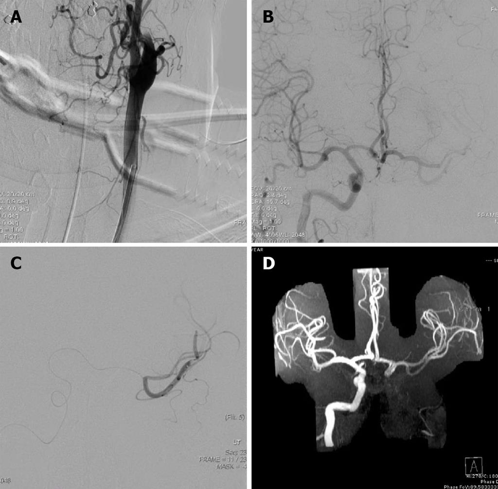
Figure 1 Pre and post intervention cerebral angiography.
A: Left carotid occlusion from likely dissection; B: Cross filling of the left internal carotid artery distribution via right internal carotid artery injection. The left middle cerebral artery (MCA) distribution does not completely opacify due to thrombus; C: Microcatheter crossing into the left MCA via the anterior communicating artery. Intra-arteria tissue plasminogen activator was delivered; D: Magnetic resonance angiography performed 10 h later confirms patency of the left MCA branches correlating with the patient’s resolution of clinical symptoms.
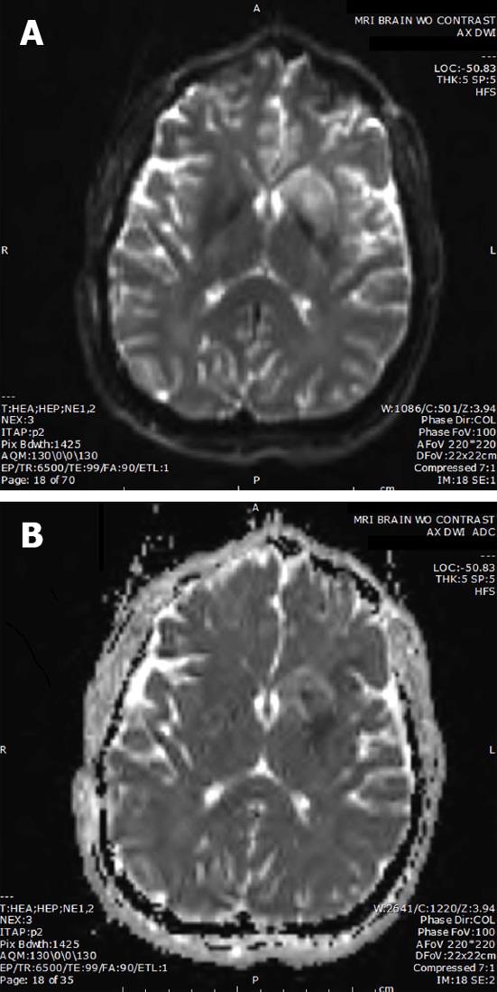
Figure 2 Ten hours post procedure imaging demonstrating small left subcortical infarct.
A: Post procedure diffusion weighted magnetic resonance imaging; B: Post procedure apparent diffusion coefficient.
-
Citation: Bulsara KR, Ediriwickrema A, Pepper J, Robertson F, Aruny J, Schindler J. Tissue plasminogen activator
via cross-collateralization for tandem internal carotid and middle cerebral artery occlusion. World J Clin Cases 2013; 1(9): 290-294 - URL: https://www.wjgnet.com/2307-8960/full/v1/i9/290.htm
- DOI: https://dx.doi.org/10.12998/wjcc.v1.i9.290


