Copyright
©2013 Baishideng Publishing Group Co.
World J Clin Cases. Dec 16, 2013; 1(9): 276-284
Published online Dec 16, 2013. doi: 10.12998/wjcc.v1.i9.276
Published online Dec 16, 2013. doi: 10.12998/wjcc.v1.i9.276
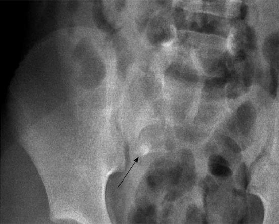
Figure 1 A nodular calcified appendicolith (arrow) in the right lower abdominal quadrant.
Figure 2 Acute appendicitis.
A: A blind-ending, non-compressible tubular structure (arrows); an echogenic appendicolith with an acoustic shadow over tip (arrowheads); B: An enlargement of whole diameter of the appendix (arrows) with enhancement and thickening of the appendiceal wall associated with intraluminal fluid collections.
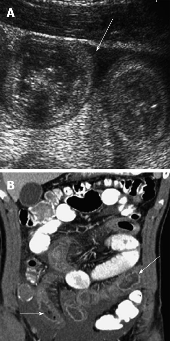
Figure 3 ‘‘Pseudokidney’’ sign (A) and ‘‘target sign (arrow)” (B) on ultrasonography.
Figure 4 A concentric ring of the ileum (arrows) from ileo-colic intussusception.
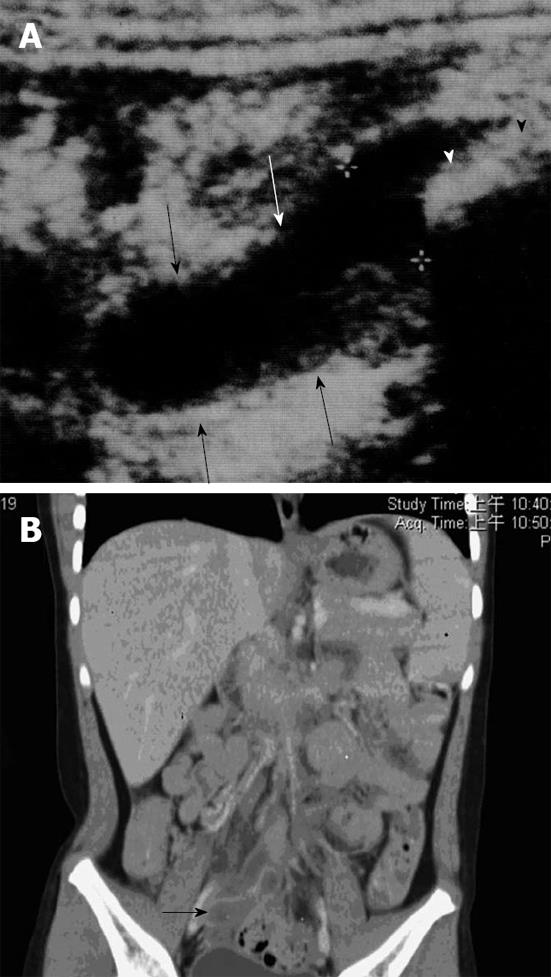
Figure 5 Henoch-Schonlein purpura.
A: Sagittal ultrasound image shows more moderate wall thickening of small bowel with ascites; B: Coronal computed tomography shows long segments of thickened, enhancing, fluid filled small bowel (arrows).
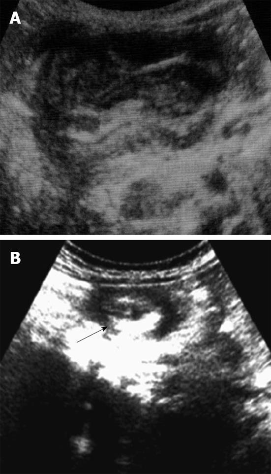
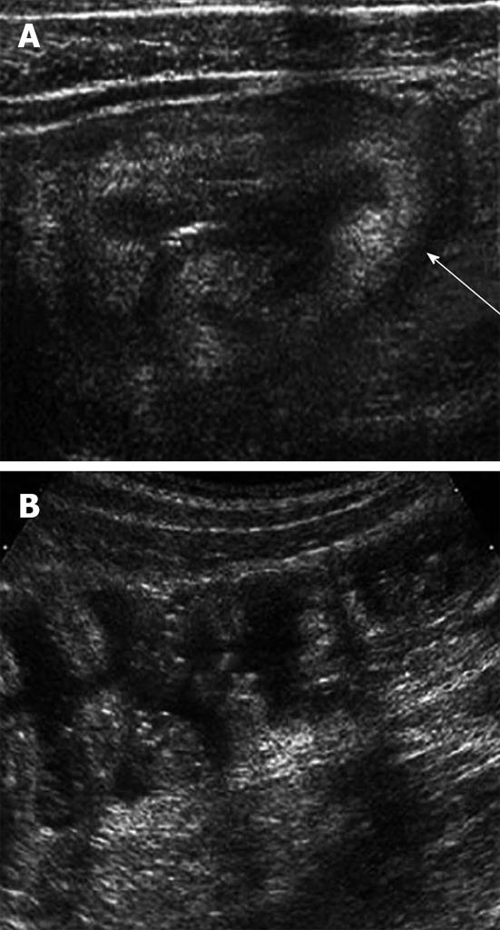
Figure 6 Bacterial enteritis.
A: Ultrasound showing marked wall thickening of the cecum (arrow) in a child with right lower quadrant pain, which returned to normal; B: 4 d later. Stool cultures were positive for enterohemorrhagic Escherichia coli.
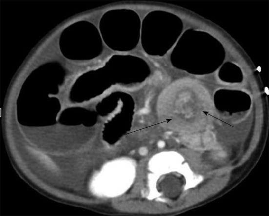
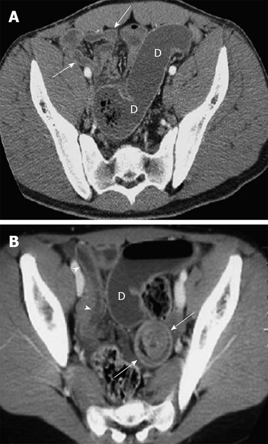
Figure 7 Meckel’s diverticulum.
A: Small-bowel obstruction shown on computed tomography (CT) in an 18-year-old boy with pathologically proven Meckel’s diverticulum; B: CT image in an 11-year-old girl shows intussusception (arrows) as a bowel loop containing alternating rings of attenuation. Note dilated proximal small bowel (D) and collapsed terminal ileum (arrowheads).
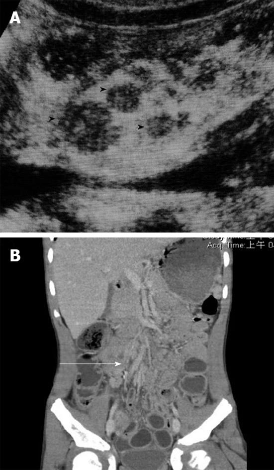
Figure 8 Mesenteric adenitis.
A: Ultrasound shows multiple enlarged lymph nodes (arrowheads) at the base of mesentery, anterior to the inferior vena cava; B: Computed tomography of the abdomen showing clustering of mesenteric lymph nodes with largest diameter of about 11.2 mm (black arrow) and thickening of the bowel wall of terminal ileum.
- Citation: Yang WC, Chen CY, Wu HP. Etiology of non-traumatic acute abdomen in pediatric emergency departments. World J Clin Cases 2013; 1(9): 276-284
- URL: https://www.wjgnet.com/2307-8960/full/v1/i9/276.htm
- DOI: https://dx.doi.org/10.12998/wjcc.v1.i9.276


