Copyright
©2013 Baishideng Publishing Group Co.
World J Clin Cases. Dec 16, 2013; 1(9): 262-275
Published online Dec 16, 2013. doi: 10.12998/wjcc.v1.i9.262
Published online Dec 16, 2013. doi: 10.12998/wjcc.v1.i9.262
Figure 1 Xp11 translocation carcinoma (hematoxylin and eosin).
A: The tumor is composed of cells with clear to eosinophilic, abundant voluminous cytoplasm (× 40); B: Psammoma bodies in stromal hyaline nodules are frequently seen, but are not required for diagnosis (× 100); C: TFE3 nuclear immunohistochemical stain can assist with confirmation of the diagnosis (× 100), but can also be positive in tumors without the molecular translocation.
Figure 2 Mucinous and tubular spindle cell carcinoma (hematoxylin and eosin).
A: The tumor has a tubular pattern on low power with mucin present between glands (× 20); B: In some areas, a spindle cell pattern is also seen (× 40); C: The tubular pattern with intervening mucin is prominent in some tumors (× 100); D: Round nuclei with prominent nucleoli are evident on higher power (× 400).
Figure 3 Multilocular cystic clear cell renal cell carcinoma (hematoxylin and eosin).
A: The tumor is composed of multiple cysts with intervening thin, fibrous septa and scattered chronic inflammatory cells (× 40); B: On higher power, the cysts are lined by a single layer of cells with clear cytoplasm (× 200); C: Low-grade nuclear features (Fuhrman nuclear grade 1) are seen and clear cells are present within the cyst wall and on the surface lining (× 400).
Figure 4 Tubulocystic carcinoma (hematoxylin and eosin).
A: Tubulocystic carcinoma, on low power, is composed of many small cystic spaces with intervening fibrous septae (× 20); B: The cysts vary in size and shape (× 40); C: On higher power, bland cuboidal and hobnail-shaped cells are identified lining the cystic spaces (× 200).
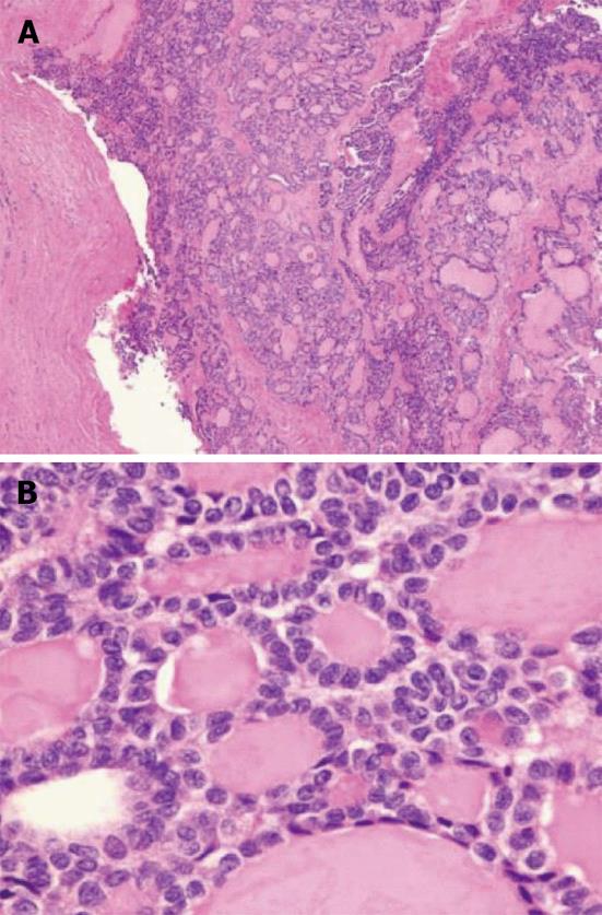
Figure 5 Thyroid-like follicular carcinoma of kidney (hematoxylin and eosin).
A: The tumor has thyroid-like follicles filled with colloid-like material on low power (× 40); B: On high power, cells with enlarged, irregular nuclei and follicular architecture, similar to the follicular neoplasms of the thyroid, are seen; Nuclear grooves are also evident (× 400). Courtesy of Dr. Pheroze Tamboli, MD Anderson Cancer Center.
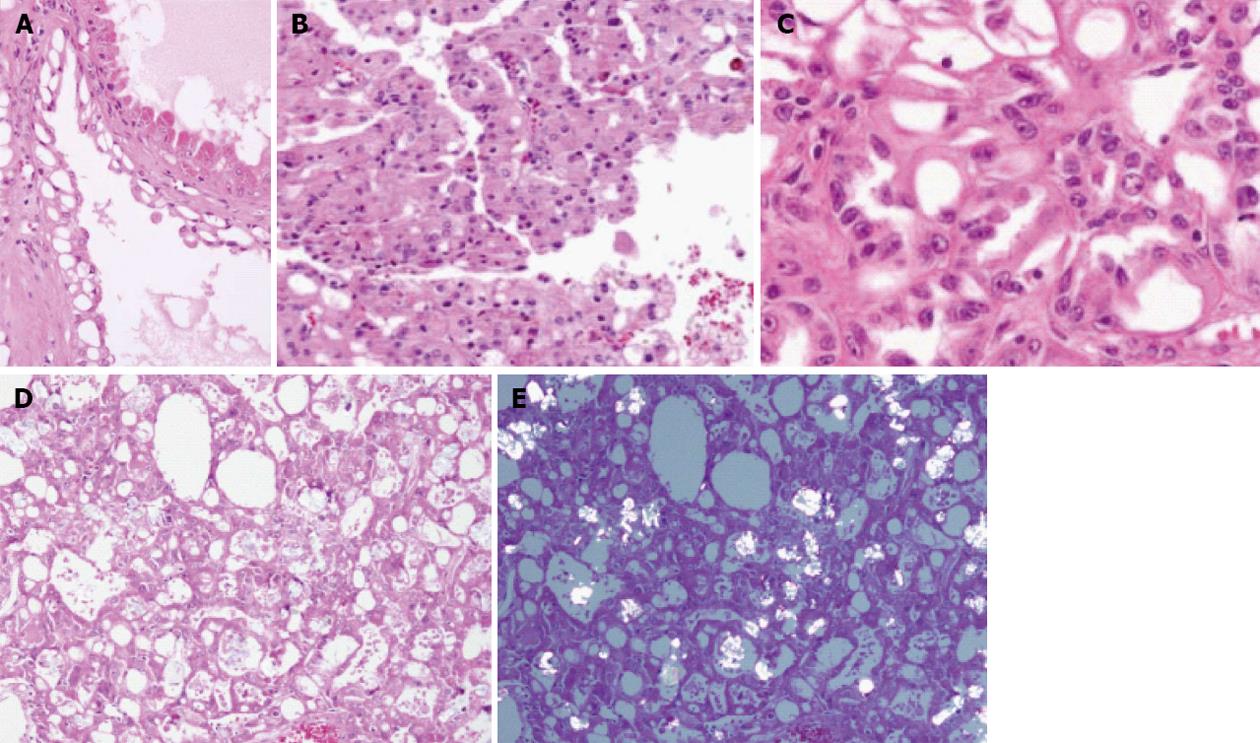
Figure 6 Acquired cystic disease-associated renal cell carcinoma.
A, B: The tumor cells in acquired cystic disease-associated renal cell carcinoma typically have abundant eosinophilic cytoplasm and are seen lining the cystic spaces (A, × 400), and sometimes also show a solid growth pattern (B, × 200); The tumor characteristically arranges in a sieve-like cribriform pattern; C: The nuclear features are consistent with a Fuhrman nuclear grade 3, with prominent nucleoli (× 600); D, E: Calcium oxalate crystals are often associated with these tumors, and can be seen on hematoxylin and eosin staining (D, × 600) and with polarization (E, × 100).
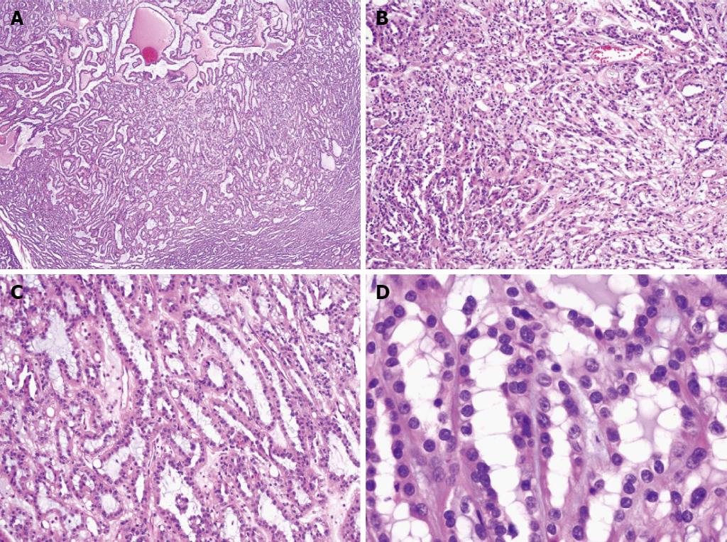
Figure 7 Clear cell papillary renal cell carcinoma (hematoxylin and eosin).
A: The tumor has both a tubular and papillary architecture within a dense fibrous stroma (× 40); B: Other areas show a tubulopapillary growth pattern with delicate fibrovascular cores and conspicuous cytoplasmic clearing (× 100); C: The characteristic clear cells with low Fuhrman nuclear grade are seen with inverted linear positioning of the nuclei away from the basement membrane (× 200).
Figure 8 Renal cell carcinoma with t(6;11) translocation (hematoxylin and eosin).
The tumor is composed of large epithelioid cells with abundant clear to eosinophilic cytoplasm; the distinctive feature of hyaline material with surrounding cells “rosette forming” is seen (× 100). Courtesy of Dr. Liang Cheng, Indiana University.
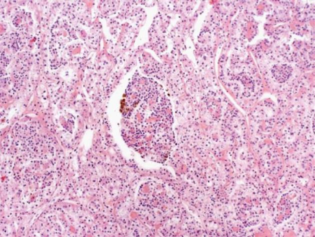
Figure 9 Hybrid oncocytoma/chromophobe renal cell carcinoma (hematoxylin and eosin).
A: Low power assessment of the tumors shows a well-circumscribed tumor with a solid growth pattern (× 40); B: The tumor cells have abundant eosinophilic cytoplasm and round, monotonous nuclei (× 200); C: Perinuclear halos are prominent throughout, with some areas showing mild nuclear irregularity (× 400). The overlapping features of oncocytoma (round nuclei with abundant eosinophilic cytoplasm) and chromophobe (perinuclear halos) are noted.
Figure 10 Renal angiomyoadenomatous tumor (hematoxylin and eosin).
A, B: The tumor is composed of epithelioid cells in a background of dense, leiomyomatous stroma (A, × 40 and B, × 100); C: The tumor cells have oval nuclei of low Fuhrman nuclear grade with clear to eosinophilic cytoplasm and protrude into the lumen, resembling a so-called “shark’s smile” (× 400). Courtesy of Dr. Melissa Stanton, Mayo Clinic, Arizona.
- Citation: Crumley SM, Divatia M, Truong L, Shen S, Ayala AG, Ro JY. Renal cell carcinoma: Evolving and emerging subtypes. World J Clin Cases 2013; 1(9): 262-275
- URL: https://www.wjgnet.com/2307-8960/full/v1/i9/262.htm
- DOI: https://dx.doi.org/10.12998/wjcc.v1.i9.262


