Copyright
©2013 Baishideng Publishing Group Co.
World J Clin Cases. Oct 16, 2013; 1(7): 227-229
Published online Oct 16, 2013. doi: 10.12998/wjcc.v1.i7.227
Published online Oct 16, 2013. doi: 10.12998/wjcc.v1.i7.227
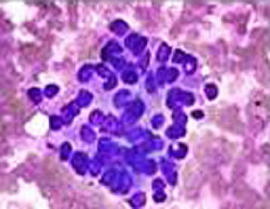
Figure 1 Fine Needle Aspiration Cytology confirmed the presence of atypical cells.
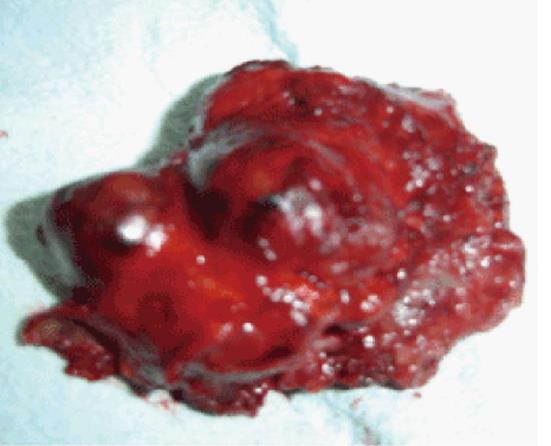
Figure 2 The cut surface was grey white and firm with focal areas of hemorrhages, necrosis.
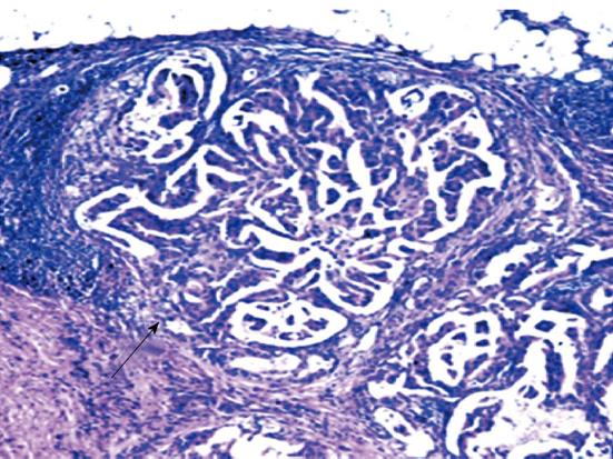
Figure 3 Showing intraductalmalignant cells arranged in papillary fronds exhibiting features of malignancy (Insitu Papillary Carcinoma of Breast), × 10.
- Citation: Ingle SB, Hinge(Ingle) CR, Murdeshwar HG, Adgaonkar BD. Unusual case of insitu (intracystic) papillary carcinoma of breast. World J Clin Cases 2013; 1(7): 227-229
- URL: https://www.wjgnet.com/2307-8960/full/v1/i7/227.htm
- DOI: https://dx.doi.org/10.12998/wjcc.v1.i7.227


