Copyright
©2013 Baishideng Publishing Group Co.
World J Clin Cases. Aug 16, 2013; 1(5): 181-186
Published online Aug 16, 2013. doi: 10.12998/wjcc.v1.i5.181
Published online Aug 16, 2013. doi: 10.12998/wjcc.v1.i5.181
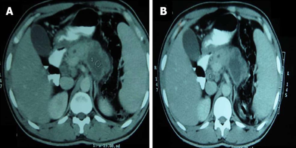
Figure 1 Contrast-enhanced computerized tomography of abdomen showing bulky pancreas (A), retro pancreatic lymphadenopathy (B) and hypodense lesion in the body and tail of the pancreas (3.
5 cm × 2.4 cm) with peripheral enhancement.
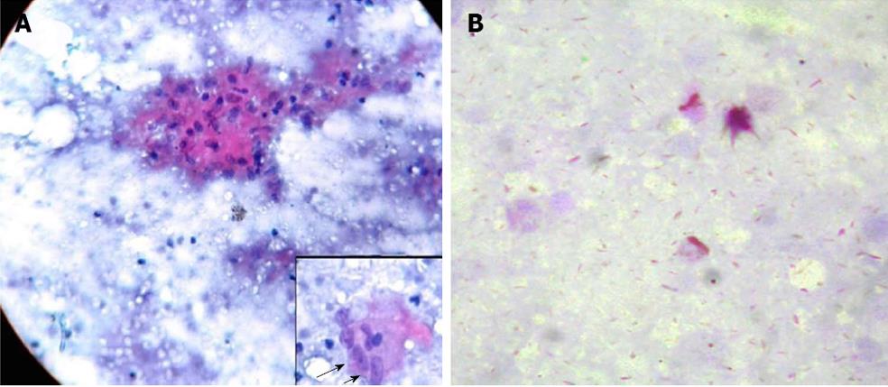
Figure 2 CT guided fine-needle aspiration cytology from the pancreatic lesion shows collection of epitheloid cells (inset) forming granuloma along with pancreatic ductal and acinar cells with patchy necrotic material (Giemsa staining) (A), Ziehl-Neelsen stain showing acid fast bacilli in the background of proteinaceous material (B).
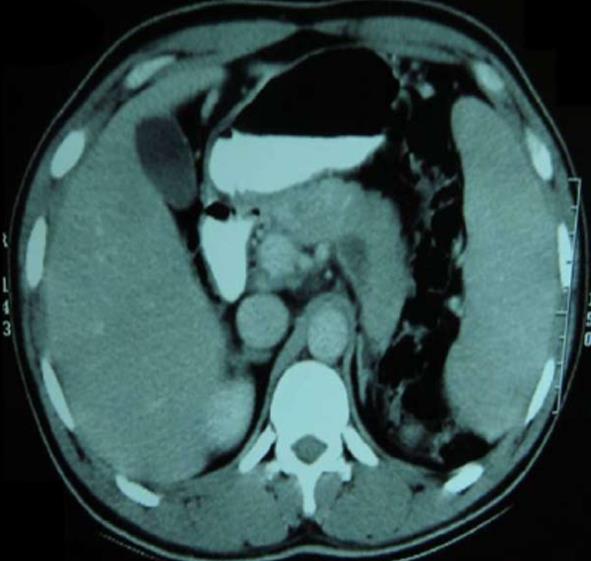
Figure 3 Review Contrast-enhanced computerized tomography of the abdomen performed at 9 mo of ATT showing near complete regression of pancreas size and resolution of hypodense lesion at body and tail.
- Citation: Sonthalia N, Ray S, Pal P, Saha A, Talukdar A. Fine needle aspiration diagnosis of isolated pancreatic tuberculosis: A case report. World J Clin Cases 2013; 1(5): 181-186
- URL: https://www.wjgnet.com/2307-8960/full/v1/i5/181.htm
- DOI: https://dx.doi.org/10.12998/wjcc.v1.i5.181


