Copyright
©2013 Baishideng Publishing Group Co.
World J Clin Cases. Aug 16, 2013; 1(5): 176-180
Published online Aug 16, 2013. doi: 10.12998/wjcc.v1.i5.176
Published online Aug 16, 2013. doi: 10.12998/wjcc.v1.i5.176
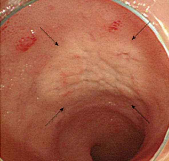
Figure 1 Color-faded cobble-stone like erosion in the ileum (arrows).
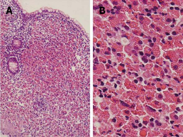
Figure 2 Biopsy specimen taken from the erosion in the ileum, showing proliferation of mononuclear cells containing numerous Russell bodies in the submucosal layer.
A: HE, × 100; B: × 400.
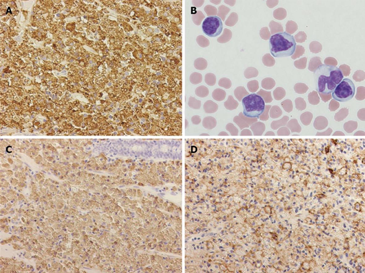
Figure 3 Immunohistochemistry.
A: Immunohistochemistry for lambda light chains using biopsy specimen from the ileum (× 400); B: Atypical lymphocytes in peripheral blood diagnosed as T-prolymphocytic leukemia (Giemsa stain, × 1000); C: Immunohistochemistry for CD79a using biopsy specimen from the ileum (× 200); D: Immunohistochemistry for CD138 using biopsy specimen from the ileum (× 200).
- Citation: Kai K, Miyahara M, Tokuda Y, Kido S, Masuda M, Takase Y, Tokunaga O. A case of mucosa-associated lymphoid tissue lymphoma of the gastrointestinal tract showing extensive plasma cell differentiation with prominent Russell bodies. World J Clin Cases 2013; 1(5): 176-180
- URL: https://www.wjgnet.com/2307-8960/full/v1/i5/176.htm
- DOI: https://dx.doi.org/10.12998/wjcc.v1.i5.176


