Copyright
©2013 Baishideng Publishing Group Co.
World J Clin Cases. Aug 16, 2013; 1(5): 172-175
Published online Aug 16, 2013. doi: 10.12998/wjcc.v1.i5.172
Published online Aug 16, 2013. doi: 10.12998/wjcc.v1.i5.172
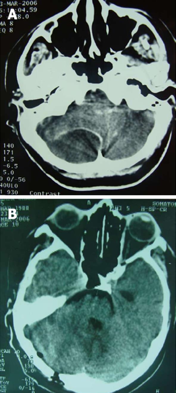
Figure 1 Computed tomography.
A: Head computed tomography showing subdural empyema of right cerebellar convexity; fourth ventricle compression and occlusion; B: Burr hole on the right occipital infratentorial convexity; reappearance of fourth ventricle; complete evacuation of subdural cerebellar empyema.
- Citation: Alimehmeti R, Seferi A, Stroni G, Sallavaci S, Rroji A, Pilika K, Petrela M. Burr hole evacuation for infratentorial subdural empyema. World J Clin Cases 2013; 1(5): 172-175
- URL: https://www.wjgnet.com/2307-8960/full/v1/i5/172.htm
- DOI: https://dx.doi.org/10.12998/wjcc.v1.i5.172


