Copyright
©2013 Baishideng Publishing Group Co.
World J Clin Cases. Aug 16, 2013; 1(5): 159-161
Published online Aug 16, 2013. doi: 10.12998/wjcc.v1.i5.159
Published online Aug 16, 2013. doi: 10.12998/wjcc.v1.i5.159
Figure 1 Ultrasonography.
A: Modified apical 4 chamber two-dimensional echocardiographic view showing dilated coronary sinus; B: Injection of agitated saline into the left antecubital vein results in filling of the coronary sinus first (star), followed by the filling of the right atrium. CS: Coronary sinus; LV: Left ventricle; RA: Right atrium; RV: Right ventricle.
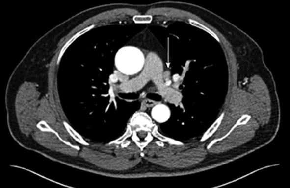
Figure 2 The axial image of cardiac computed tomography angiography shows the persistent left superior vena cava (arrow).
- Citation: Siddiqui AM, Cao LB, Movahed A. Side matters: An intriguing case of persistent left superior vena-cava. World J Clin Cases 2013; 1(5): 159-161
- URL: https://www.wjgnet.com/2307-8960/full/v1/i5/159.htm
- DOI: https://dx.doi.org/10.12998/wjcc.v1.i5.159


