Copyright
©2013 Baishideng Publishing Group Co.
World J Clin Cases. Jun 16, 2013; 1(3): 124-127
Published online Jun 16, 2013. doi: 10.12998/wjcc.v1.i3.124
Published online Jun 16, 2013. doi: 10.12998/wjcc.v1.i3.124
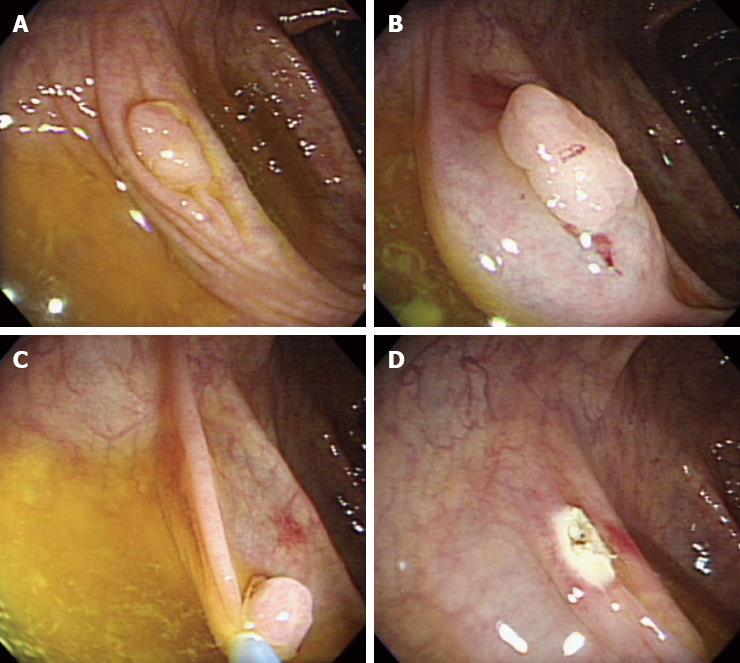
Figure 1 Colonoscopy showing a sessile polyp located in the ascending colon (A), after saline was injected (B), endoscopic mucosal resection was performed (C and D).
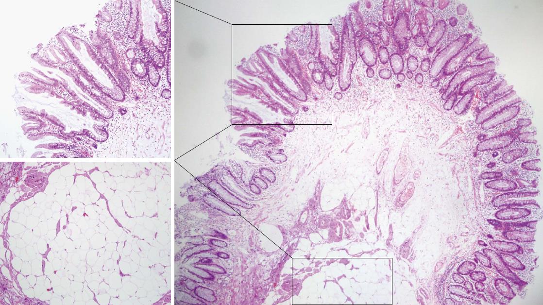
Figure 2 Microscopic image of the resected specimen showing a colonic lipoma with overlying hyperplastic epithelium in the low-magnified image (hematoxylin-eosin, × 40).
The lining epithelium resembles a hyperplastic polyp and a tumor composed of adipose tissue in the high-magnified image (hematoxylin-eosin, × 100).
- Citation: Yeom JO, Kim SY, Jang EC, Yu JY, Chang ED, Cho YS. Colonic lipoma covered by hyperplastic epithelium: Case report. World J Clin Cases 2013; 1(3): 124-127
- URL: https://www.wjgnet.com/2307-8960/full/v1/i3/124.htm
- DOI: https://dx.doi.org/10.12998/wjcc.v1.i3.124


