Copyright
©2013 Baishideng Publishing Group Co.
World J Clin Cases. Jun 16, 2013; 1(3): 121-123
Published online Jun 16, 2013. doi: 10.12998/wjcc.v1.i3.121
Published online Jun 16, 2013. doi: 10.12998/wjcc.v1.i3.121
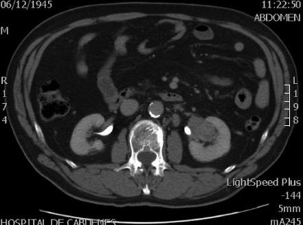
Figure 1 Abdominal contrast enhanced tomography with left kidney
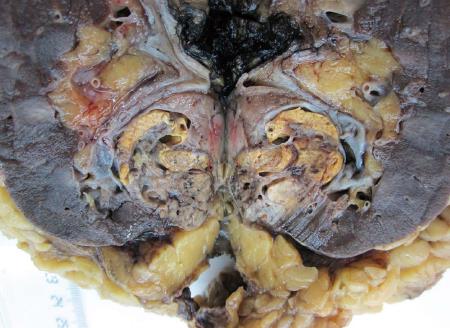
Figure 2 Yellow kidney tumor in touch with rose node, located at hilum, 4 cm in diameter.
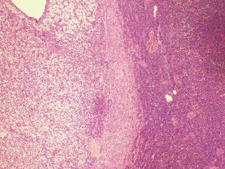
Figure 3 A microscopic view of the tumors.
In the left, renal conventional clear cell carcinoma. In the right, a lymph node with small B cell lymphoma.
- Citation: Fernandez-Pello S, Rodriguez Villamil L, Gonzalez Rodriguez I, Venta V, Cuervo J, Menéndez CL. Lymph node non-Hodgkin's lymphoma incidentally discovered during a nephrectomy for renal cell carcinoma. World J Clin Cases 2013; 1(3): 121-123
- URL: https://www.wjgnet.com/2307-8960/full/v1/i3/121.htm
- DOI: https://dx.doi.org/10.12998/wjcc.v1.i3.121


