Copyright
©2013 Baishideng Publishing Group Co.
World J Clin Cases. Jun 16, 2013; 1(3): 116-120
Published online Jun 16, 2013. doi: 10.12998/wjcc.v1.i3.116
Published online Jun 16, 2013. doi: 10.12998/wjcc.v1.i3.116
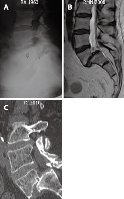
Figure 1 Seriated radiological exams showing the progression of the spondylolisthesis from grade I in 1963, to grade III in 2010.
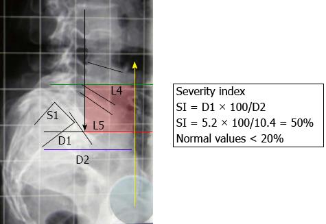
Figure 2 Analysis of the severity index and of the square of unstable zone.
It’s described by Lamartina[14]. SI: Severity index.
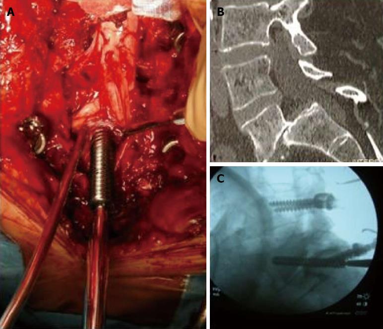
Figure 3 Intraoperative picture that showed the insertion of the trans sacral screw.
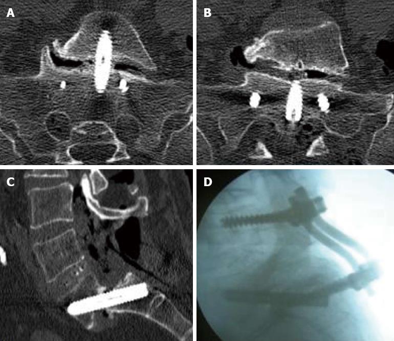
Figure 4 Computer tomography scan at 12 mo follow-up that showed the correct positioning and fusion of the system.
- Citation: Landi A, Marotta N, Mancarella C, Tarantino R, Delfini R. Trans-sacral screw fixation in the treatment of high dyplastic developmental spondylolisthesis. World J Clin Cases 2013; 1(3): 116-120
- URL: https://www.wjgnet.com/2307-8960/full/v1/i3/116.htm
- DOI: https://dx.doi.org/10.12998/wjcc.v1.i3.116


