Copyright
©2013 Baishideng.
World J Clin Cases. Apr 16, 2013; 1(1): 59-63
Published online Apr 16, 2013. doi: 10.12998/wjcc.v1.i1.59
Published online Apr 16, 2013. doi: 10.12998/wjcc.v1.i1.59
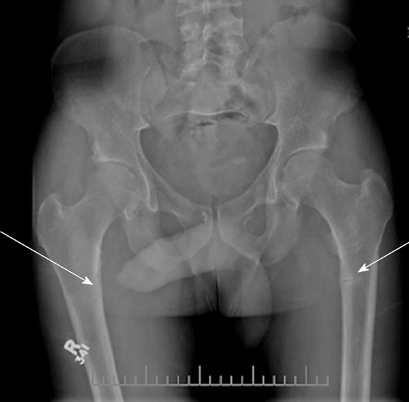
Figure 1 Bilateral radiographs of femurs.
Arrows indicate Looser’s zones.
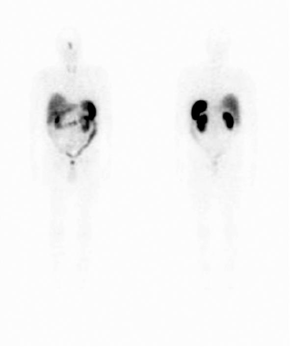
Figure 2 Octreotide scan demonstrating uptake in the sinus area.
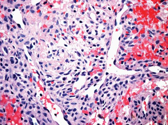
Figure 3 Photomicrograph demonstrating spindle cell neoplasm with prominent thin-walled branching vasculature.
The tumour is comprised of short fascicles of cells with pale eosinophilic cytoplasm and bland ovoid nuclei. There is conspicuous extravasation of erythrocytes (haematoxylin and eosin, × 200).
- Citation: Jamal SA, Dickson BC, Radziunas I. Tumour induced osteomalacia due to a sinonasal hemangiopericytoma: A case report. World J Clin Cases 2013; 1(1): 59-63
- URL: https://www.wjgnet.com/2307-8960/full/v1/i1/59.htm
- DOI: https://dx.doi.org/10.12998/wjcc.v1.i1.59


