Copyright
©2013 Baishideng.
World J Clin Cases. Apr 16, 2013; 1(1): 28-30
Published online Apr 16, 2013. doi: 10.12998/wjcc.v1.i1.28
Published online Apr 16, 2013. doi: 10.12998/wjcc.v1.i1.28
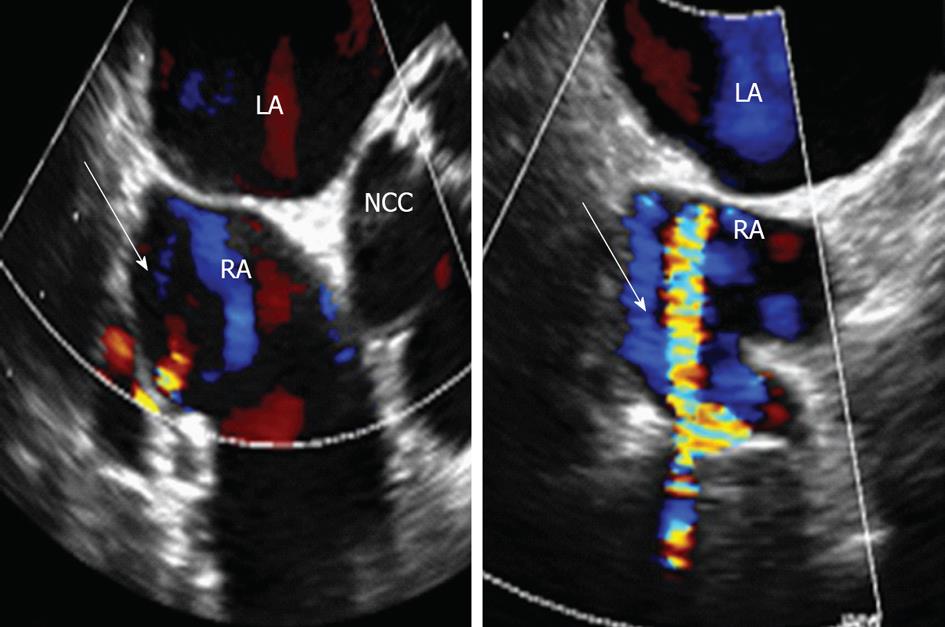
Figure 1 Transesophageal echocardiogram-mid esophageal view, short axis, at -30 degree view, with slight clockwise rotation.
White arrow points to the abnormal vascular communication noted to the right atrium on transesophageal echocardiogram. LA: Left atrium; RA: Right atrium; NCC: Noncoronary cusp.
Figure 2 Magnetic resonance angiography-transverse view of the heart with visualization of all four chambers.
White arrow points to the flow anomaly, which was really the graft coming up and running adjacent to the coronary sinus. RV: Right ventricle; LV: Left ventricle; LA: Left atrium; RA: Right atrium.
- Citation: Cao L, Farooqui MA, Wood W, Cahill J, Movahed A. Saphenous graft on transesophageal echocardiogram masquerading as an abnormal vascular communication into the right atrium. World J Clin Cases 2013; 1(1): 28-30
- URL: https://www.wjgnet.com/2307-8960/full/v1/i1/28.htm
- DOI: https://dx.doi.org/10.12998/wjcc.v1.i1.28


