Copyright
©2013 Baishideng.
World J Clin Cases. Apr 16, 2013; 1(1): 19-24
Published online Apr 16, 2013. doi: 10.12998/wjcc.v1.i1.19
Published online Apr 16, 2013. doi: 10.12998/wjcc.v1.i1.19
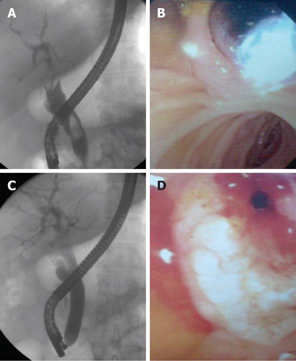
Figure 1 Endoscopic papillary large balloon dilation performed in a 58-year-old female.
A: Cholangiogram shows a large dilated common bile duct with two stones of about 2 cm each; B: Endoscopic view of the inflated balloon which it is located across the papilla after minimal endoscopic sphincterotomy; C: Fluoroscopic image of balloon dilation (16 mm diameter); D: Large biliary orifice after the procedure.
- Citation: Zippi M, De Felici I, Pica R, Traversa G, Occhigrossi G. Endoscopic papillary balloon dilation for difficult common bile duct stones: Our experience. World J Clin Cases 2013; 1(1): 19-24
- URL: https://www.wjgnet.com/2307-8960/full/v1/i1/19.htm
- DOI: https://dx.doi.org/10.12998/wjcc.v1.i1.19


