Copyright
©The Author(s) 2025.
World J Methodol. Mar 20, 2025; 15(1): 96923
Published online Mar 20, 2025. doi: 10.5662/wjm.v15.i1.96923
Published online Mar 20, 2025. doi: 10.5662/wjm.v15.i1.96923
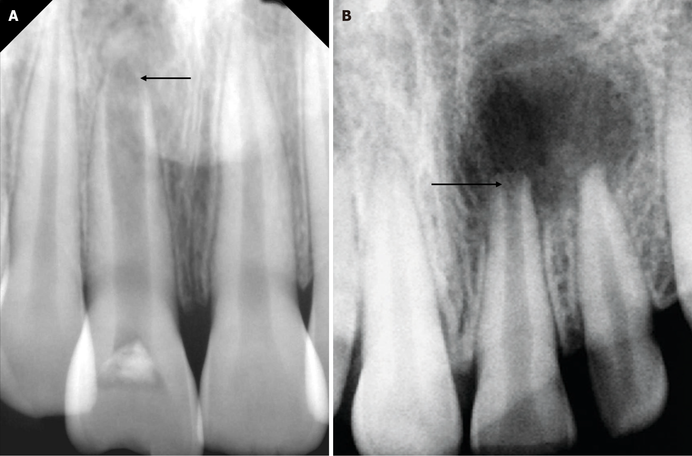
Figure 1 Two types of open apex.
A: Blunderbuss apex; B: Non-blunderbuss apex.
Figure 2 Wein’s classification.
A: No root or bone resorption is evident, and the preparation is terminated at 1.0 mm from the apical foramen; B: Bone resorption is apparent but no root resorption, and the preparation is terminated at 1.5 mm from the apical foramen; C: Both root and bone resorption are apparent, and the preparation is terminated at 2.0 mm from the apical foramen.
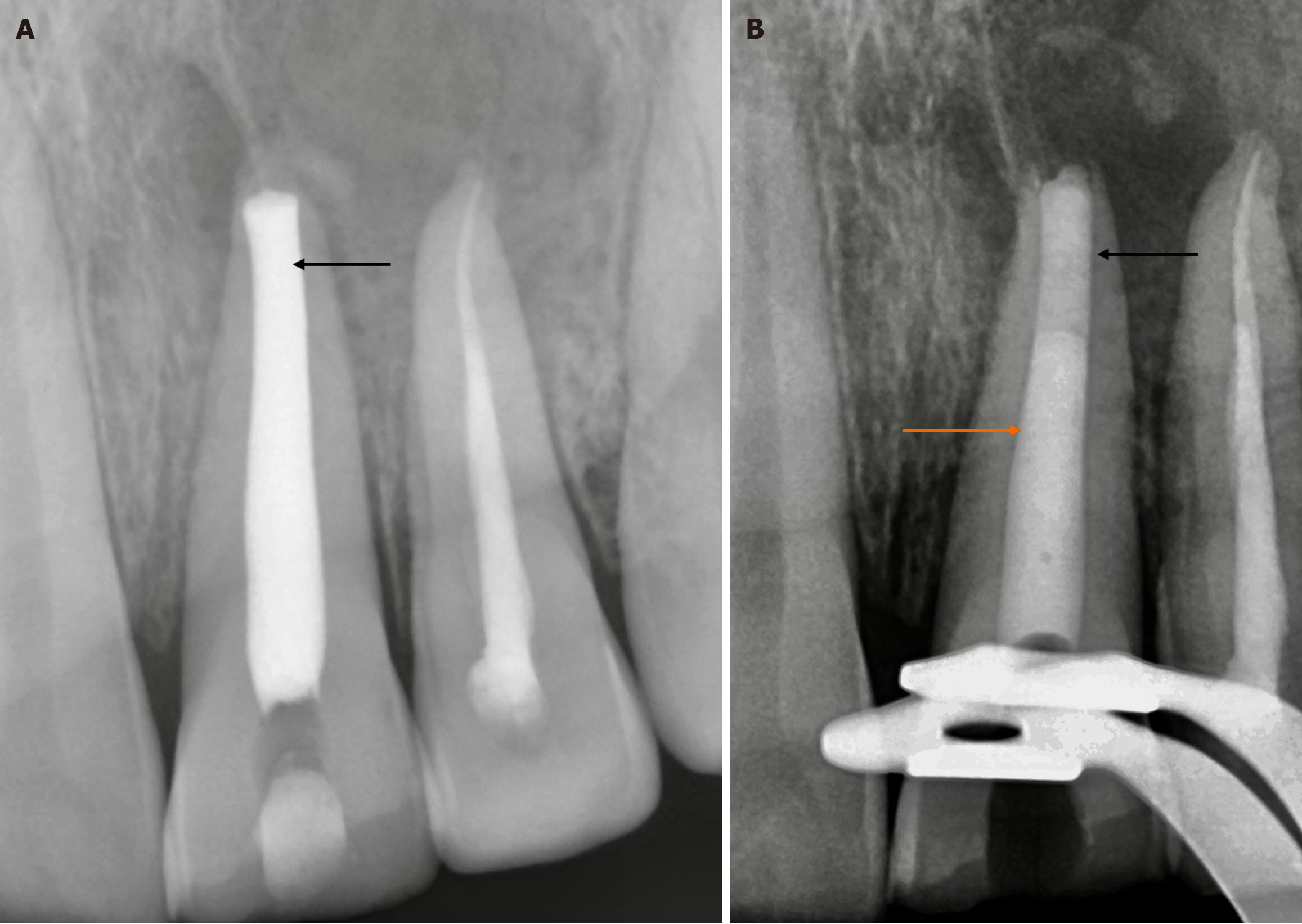
Figure 3 Different types of materials for open apex management.
A: Placement of calcium hydroxide dressing; B: Black arrow showing 5 mm mineral trioxide aggregate cement at the apex and orange arrow showing the obturation of the remaining canal.
Figure 4 Showing clinical images of the open apex and its management under the dental operating microscope.
A: Open apex at 25X under magnification; B: Placement of mineral trioxide aggregate increment at 16X under magnification.
- Citation: Chauhan S, Chauhan R, Bhasin P, Sharaf BG. Present status and future directions: Apexification. World J Methodol 2025; 15(1): 96923
- URL: https://www.wjgnet.com/2222-0682/full/v15/i1/96923.htm
- DOI: https://dx.doi.org/10.5662/wjm.v15.i1.96923


