Copyright
©The Author(s) 2015.
World J Virology. Aug 12, 2015; 4(3): 285-294
Published online Aug 12, 2015. doi: 10.5501/wjv.v4.i3.285
Published online Aug 12, 2015. doi: 10.5501/wjv.v4.i3.285
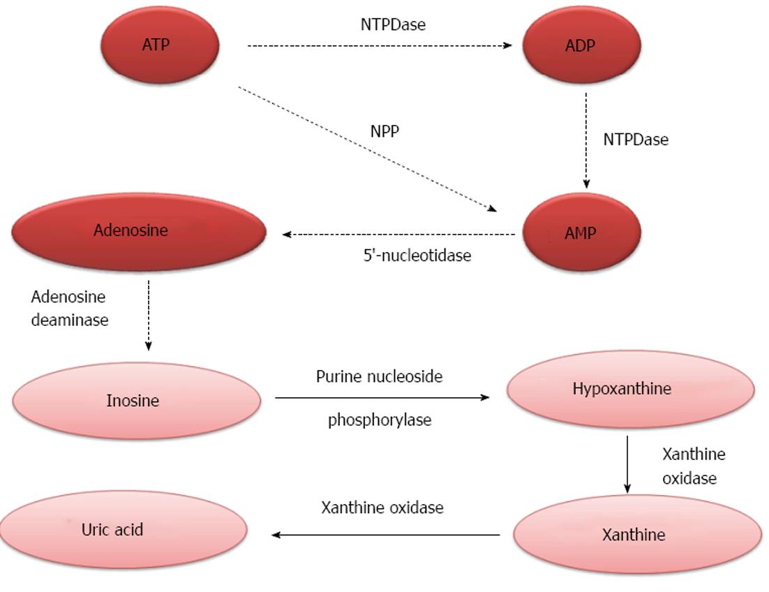
Figure 1 Schematic representation of the purinergic pathway.
ATP is broken down to ADP and AMP by NTPDase or directly to AMP by pyrophosphatase/phosphodiesterase (NPP). AMP is converted to adenosine by 5′-ectonucleotidase (CD73). Adenosine deaminase (ADA) transforms adenosine into inosine which is converted in hypoxanthine by purine nucleoside phosphorylase (PNP). Xantine oxidase catalyzes the oxidation of hypoxanthine to xanthine and xanthine to uric acid.
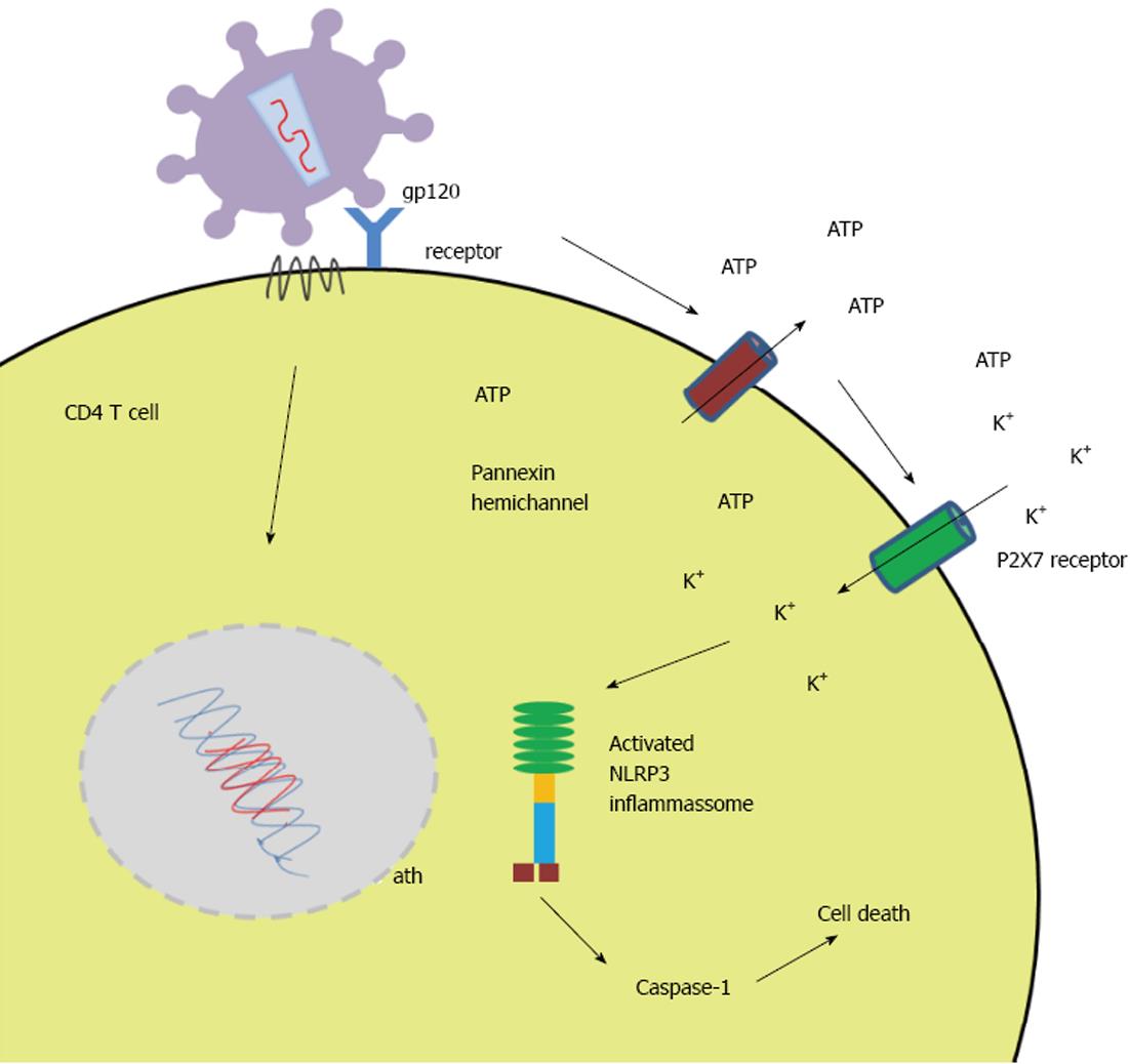
Figure 2 Schematic illustration of ATP release and activation of NOD-like receptor family, pyrin domain containing 3 inflamassome in a human immunodeficiency virus infected CD4 T cell.
Once human immunodeficiency virus gp120 binds to its receptor ATP release is stimulated through pannexin hemichannels with consequent activation of P2X7 receptor. The influx of potassium causes the activation of NOD-like receptor family, pyrin domain containing 3 (NLRP3) inflammassome leading to cell death via Caspase-1.
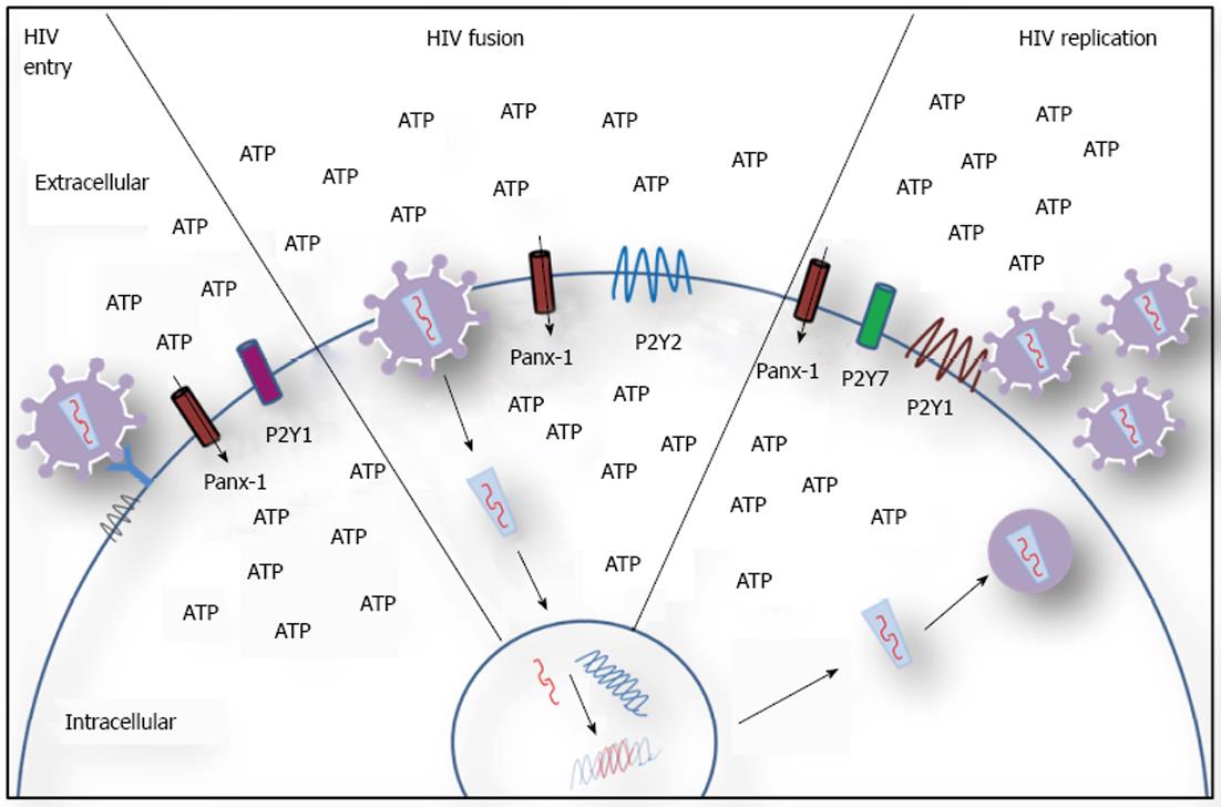
Figure 3 Schematic representation of purinergic receptors involvement in human immunodeficiency virus infection.
Pannexin hemichannels (Panx-1) are open in response to ATP release and activation of purinergic receptors, facilitating viral entry, fusion and replication. The blockage of viral entry by P2X1 antagonists suggests it is involved in this stage of infection. P2Y2 increases cell membrane depolarization facilitating fusion. P2X7 and P2Y1 are involved in later steps of viral cycle.
- Citation: Passos DF, Schetinger MRC, Leal DB. Purinergic signaling and human immunodeficiency virus/acquired immune deficiency syndrome: From viral entry to therapy. World J Virology 2015; 4(3): 285-294
- URL: https://www.wjgnet.com/2220-3249/full/v4/i3/285.htm
- DOI: https://dx.doi.org/10.5501/wjv.v4.i3.285


