Copyright
©The Author(s) 2025.
World J Virol. Mar 25, 2025; 14(1): 100003
Published online Mar 25, 2025. doi: 10.5501/wjv.v14.i1.100003
Published online Mar 25, 2025. doi: 10.5501/wjv.v14.i1.100003
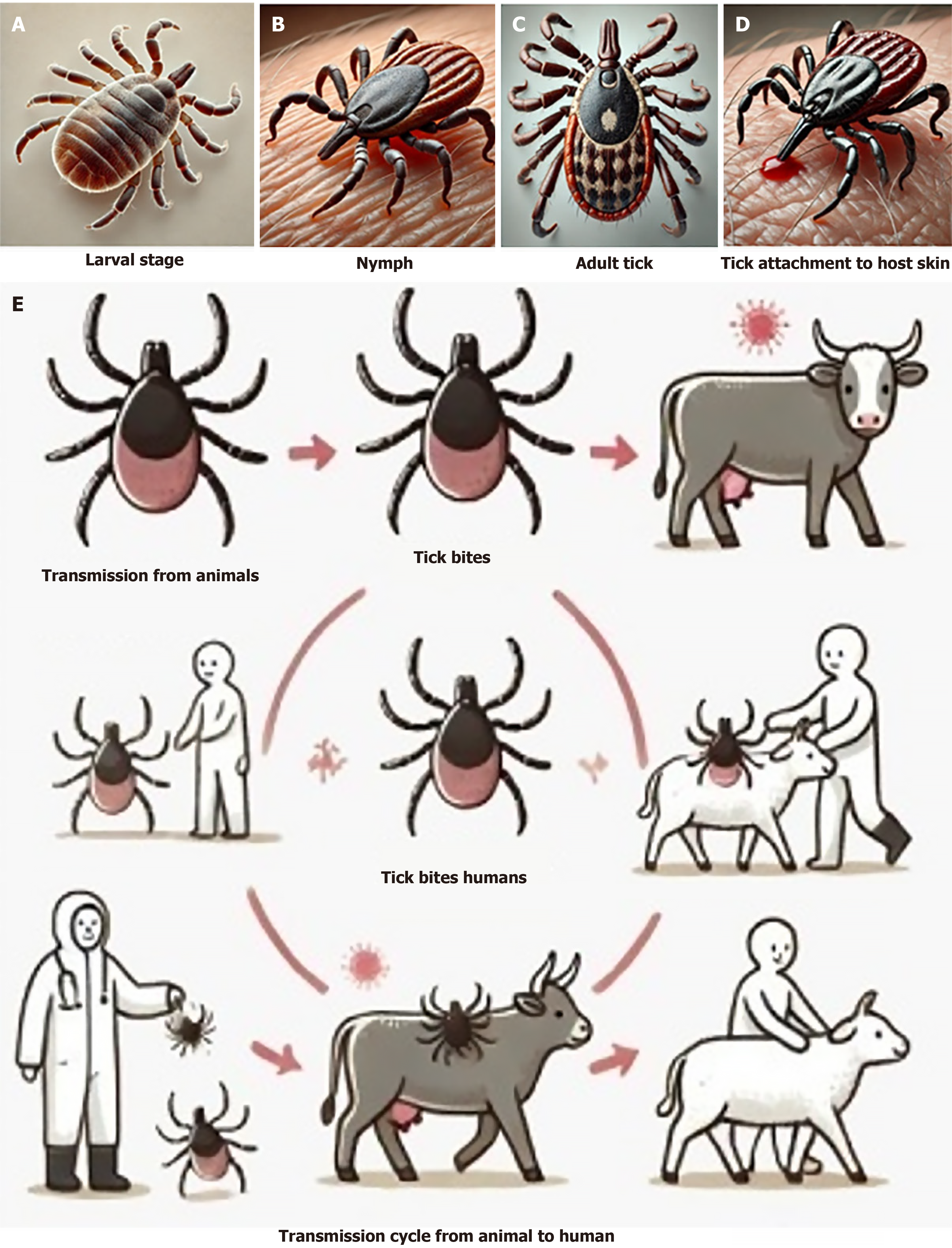
Figure 1 Lifecycle and vector role of Hyalomma ticks in Crimean-Congo hemorrhagic fever transmission.
A.Larval stage of the Hyalomma tick; B: Nymphal stage attached to a host; C: Adult Hyalomma tick, the main vector for Crimean-Congo hemorrhagic fever virus; D: Close-up of the tick attachment to host skin for blood-feeding; E: Transmission cycle from animal to human through tick bites and handling infected animals.
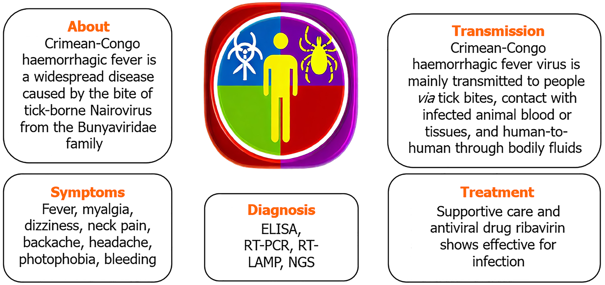
Figure 2 Overview of clinical stages and symptoms in Crimean-Congo hemorrhagic fever patients.
Early stage with fever, myalgia, and fatigue. Pre-hemorrhagic stage with signs of conjunctival hemorrhage. Hemorrhagic stage showing extensive petechiae and ecchymosis. Recovery phase after supportive treatment. Post-recovery convalescence with residual symptoms. ELISA: Enzyme-linked immunosorbent assay; RT-LAMP: Reverse transcription loop-mediated isothermal amplification; NGS: Next-generation sequencing.
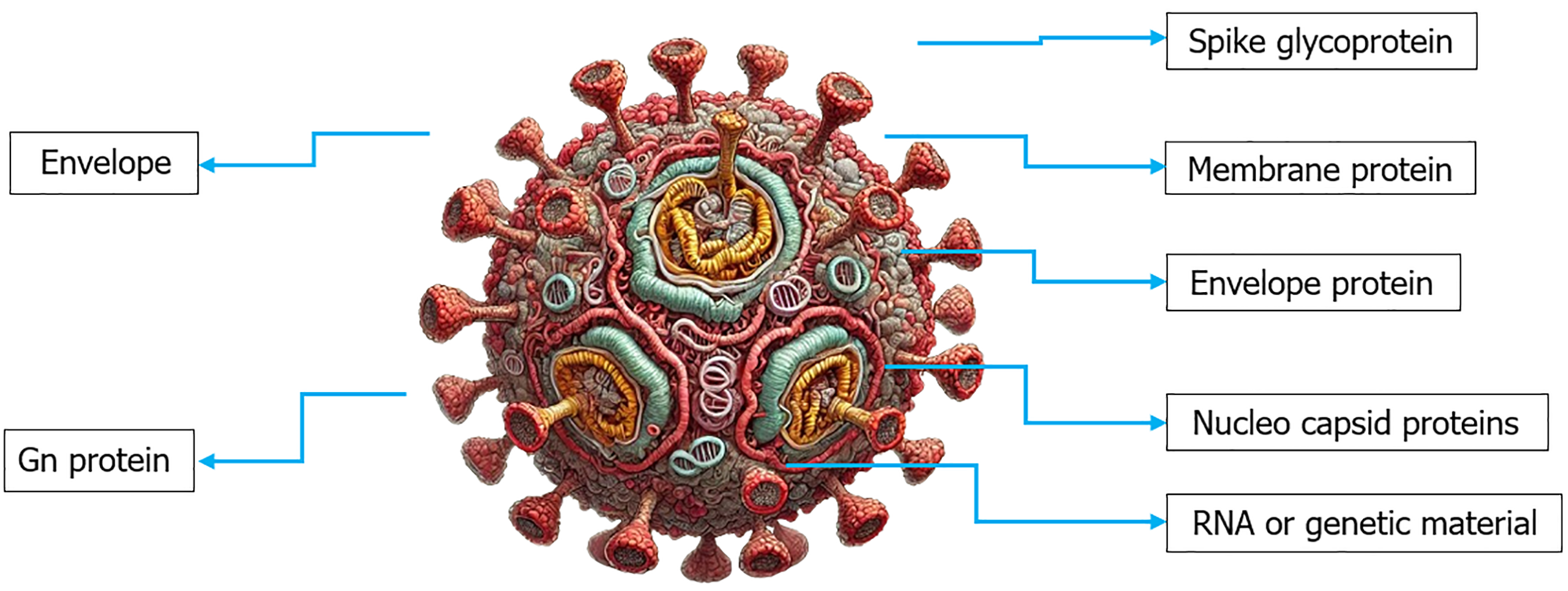
Figure 3 Structure of Crimean-Congo hemorrhagic fever virus.
This includes electron microscopy image of the Crimean-Congo hemorrhagic fever virus virion, schematic of the viral genome, showing S, M, and L RNA segments, envelope structure with Gn and Gc glycoproteins, viral RNA polymerase encoded by the L segment, nucleocapsid protein (N) from the S segment.
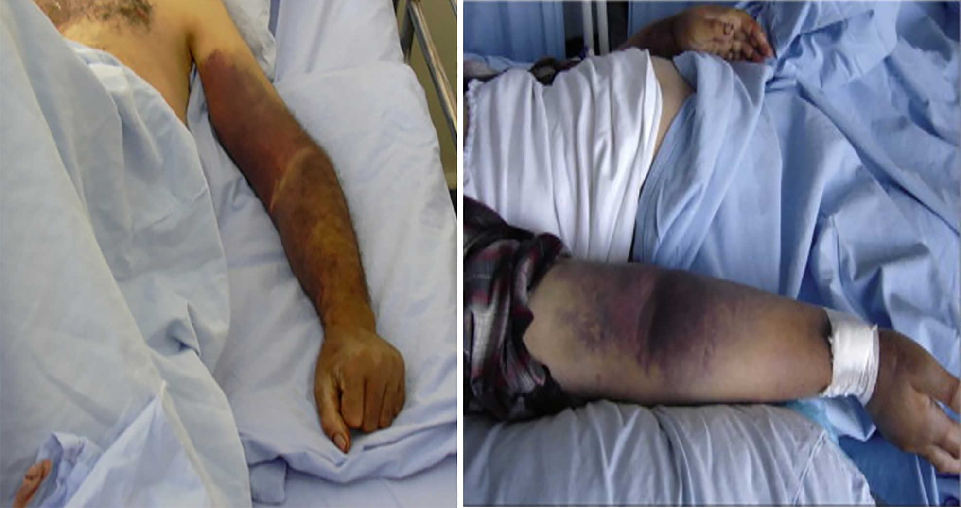
Figure 4 Petechiae on the hand of a Crimean-Congo hemorrhagic fever patient.
Petechiae are small red or purple spots resulting from capillary bleeding under the skin, commonly seen in the hemorrhagic phase of Crimean-Congo hemorrhagic fever. These non-blanching spots, distributed unevenly across the skin, indicate the severity of infection and can be accompanied by additional hemorrhagic symptoms like ecchymosis and mucosal bleeding.
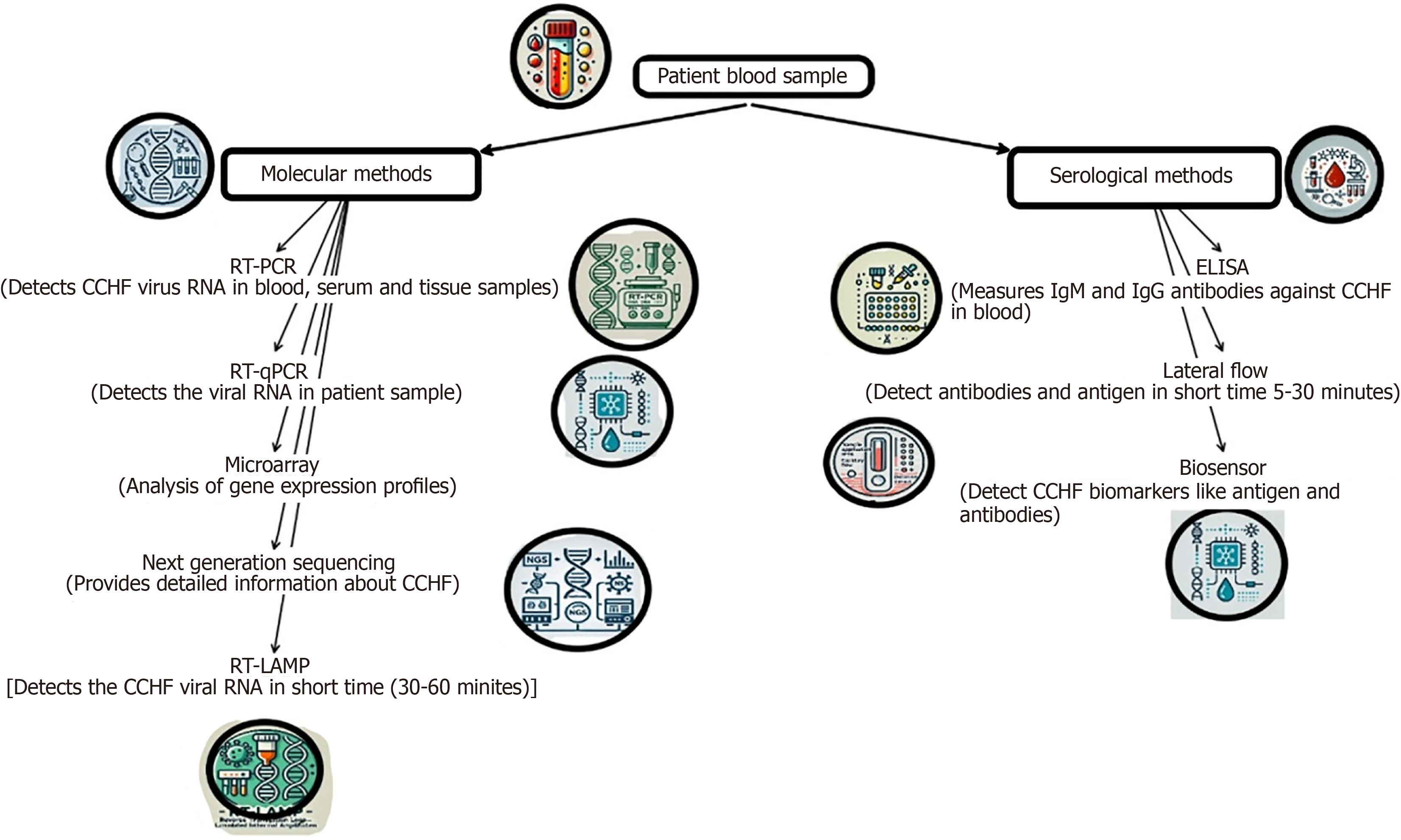
Figure 5 Laboratory diagnostic methods for Crimean-Congo hemorrhagic fever detection.
PCR amplification of Crimean-Congo hemorrhagic fever virus (CCHFV) RNA in patient blood samples, enzyme-linked immunosorbent assay antigen capture method for viral antigen detection in serum samples, viral isolation technique from infected tick tissues, immunohistochemical staining of CCHFV antigens in formalin-fixed tissue, real-time PCR results showing viral load quantification. CCHF: Crimean-Congo hemorrhagic fever; RT-LAMP: Reverse transcription loop-mediated isothermal amplification.
- Citation: Karanam SK, Nagvishnu K, Uppala PK, Edhi S, Varri SR. Crimean-Congo hemorrhagic fever: Pathogenesis, transmission and public health challenges. World J Virol 2025; 14(1): 100003
- URL: https://www.wjgnet.com/2220-3249/full/v14/i1/100003.htm
- DOI: https://dx.doi.org/10.5501/wjv.v14.i1.100003


