Copyright
©The Author(s) 2025.
World J Transplant. Sep 18, 2025; 15(3): 102798
Published online Sep 18, 2025. doi: 10.5500/wjt.v15.i3.102798
Published online Sep 18, 2025. doi: 10.5500/wjt.v15.i3.102798
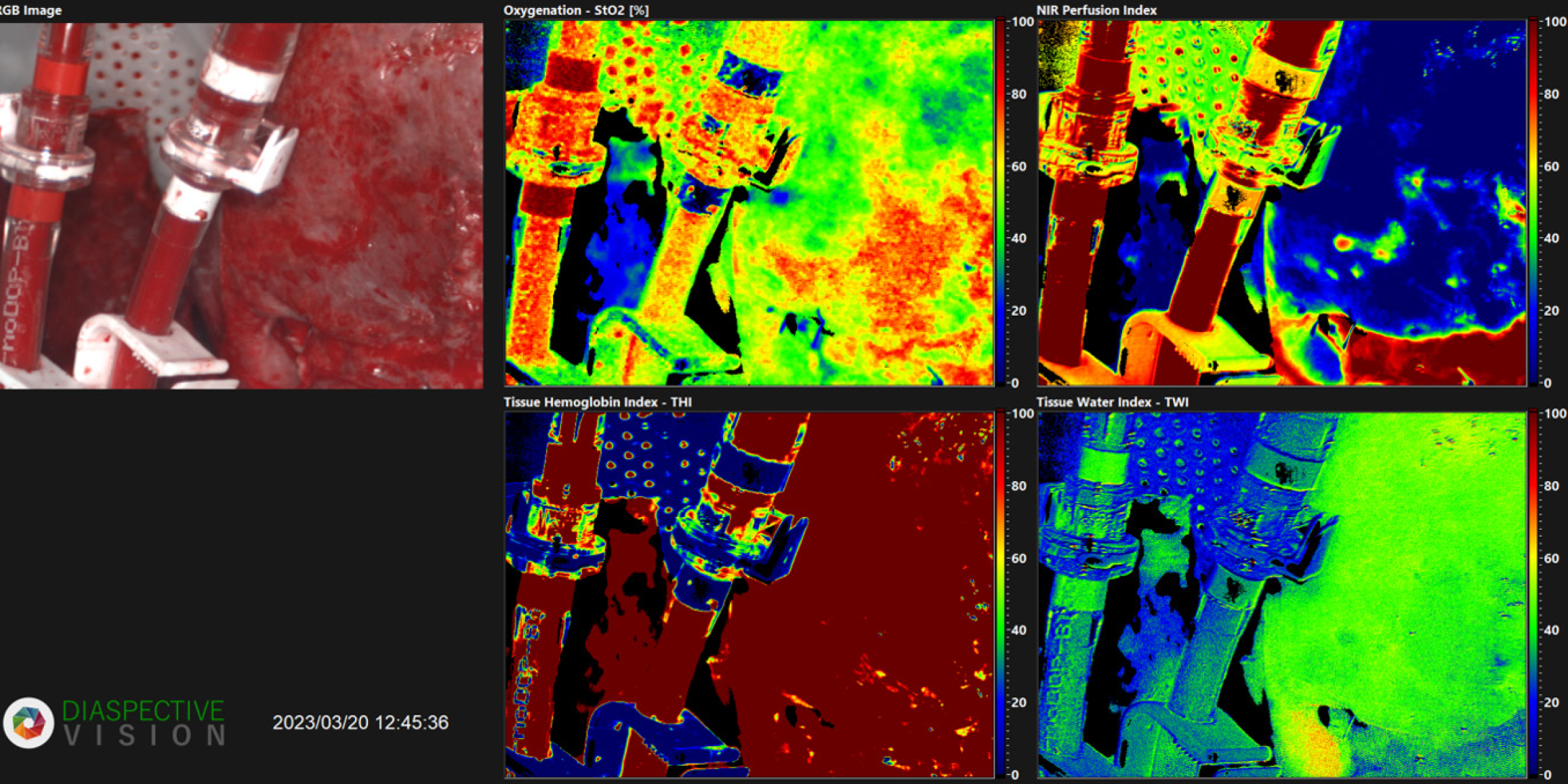
Figure 1 Hyperspectral image before discarding the liver - segments II and III.
Macroscopically, patchy alterations are observed. Hyperspectral imaging reveals decreased oxygenation on the liver surface, primarily reflected in shades of green, indicating approximately 40%-50% oxygen saturation. Moreover, near-infrared perfusion detects critically low oxygen saturation levels, ranging from 0%-20%, at a depth of 4-6 mm in those areas.
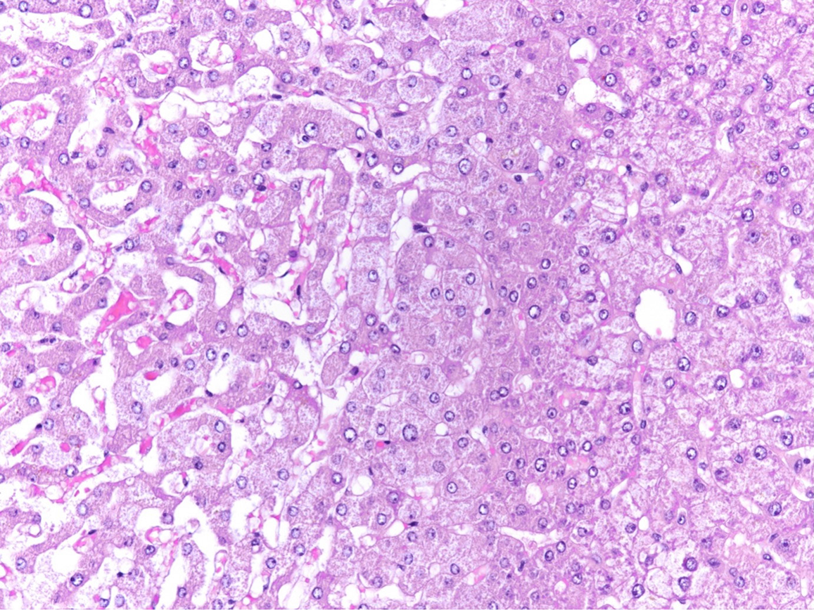
Figure 2 Histological analysis of discarded liver.
Histology shows areas of sinusoidal dilatation and congestion with atrophy and hypoxic injury of hepatocytes (left), and normal liver for comparison (right, original × 100).
- Citation: El-Mahrouk M, Langner C, Sucher R, Kniepeiss D. Introducing hyperspectral imaging as a novel tool for assessing donor liver quality during machine perfusion: A case report. World J Transplant 2025; 15(3): 102798
- URL: https://www.wjgnet.com/2220-3230/full/v15/i3/102798.htm
- DOI: https://dx.doi.org/10.5500/wjt.v15.i3.102798


