Copyright
©The Author(s) 2024.
World J Transplant. Jun 18, 2024; 14(2): 92137
Published online Jun 18, 2024. doi: 10.5500/wjt.v14.i2.92137
Published online Jun 18, 2024. doi: 10.5500/wjt.v14.i2.92137
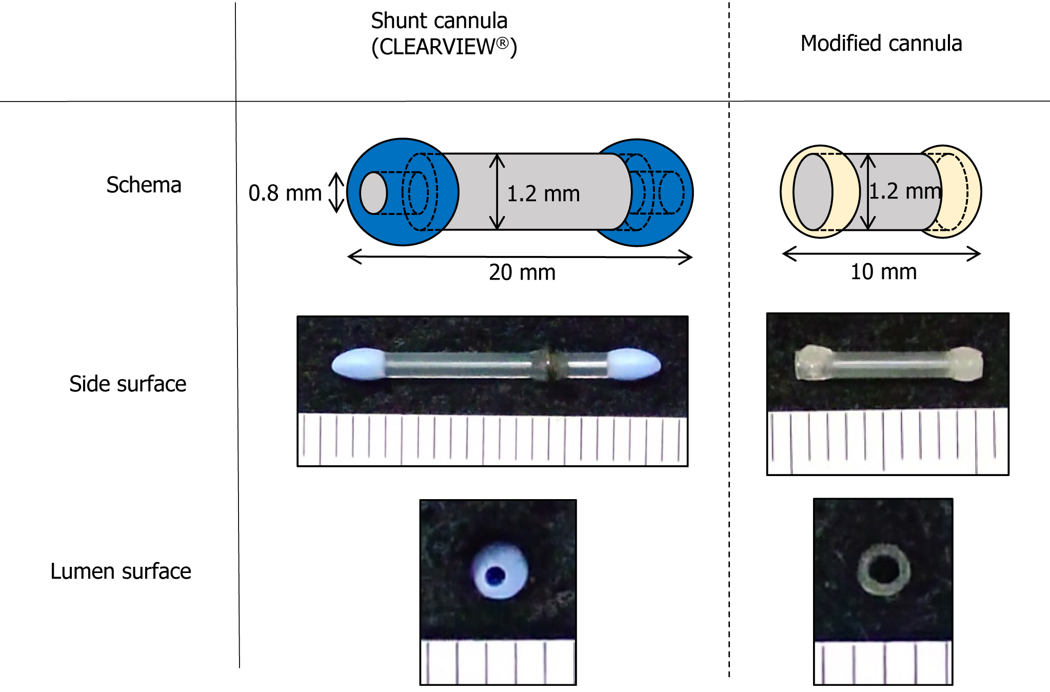
Figure 1 Shunt cannula and modified cannula.
The shunt cannula (Clearview®) is 20 mm long with a lumen diameter of 0.8 mm at the entrance and 1.2 mm at the middle. The modified cannula was 10 mm long with a lumen diameter of 1.2 mm over its entire length.
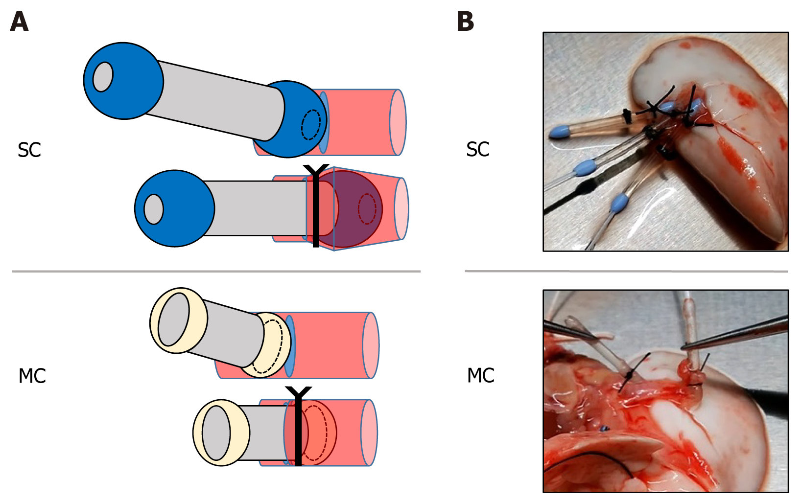
Figure 2 Graft preparation.
A: Schema of cannula insertion. The tip makes it easy to insert the cannula into the lumen and prevents the cannula from falling out during ligature fixation. Regarding the modified cannula, the tip is smaller than that of the shunt cannula, resulting in easier insertion and simpler technique of ligation and fixation is simpler; B: Left lung graft after cannula insertion. MC: Modified cannula; SC: Shunt cannula.
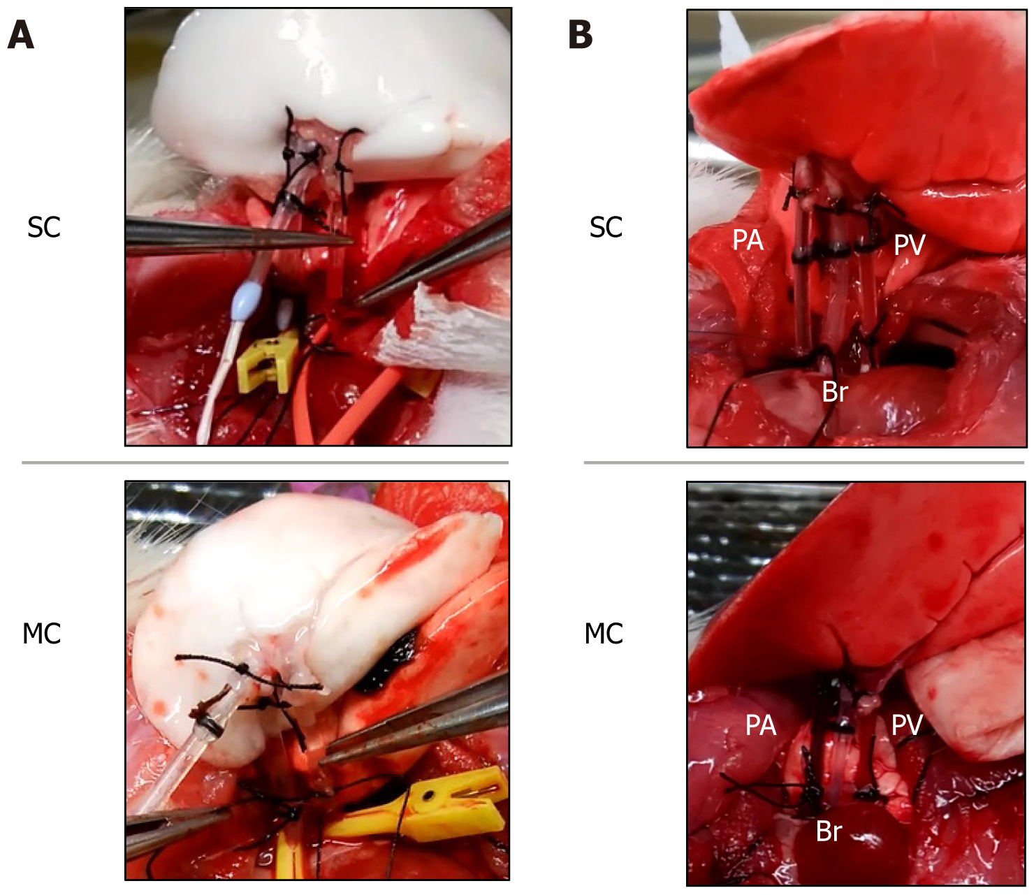
Figure 3 Recipient operation.
A: Pulmonary vein anastomosis using a cannula; B: After lung graft revascularization, venous blood perfusion is visible in the pulmonary artery, and arterial blood perfusion is visible in the pulmonary artery. In the modified cannula group, the grafted lung is located closer to the pulmonary hilum compared with the shunt cannul group. PA: Pulmonary artery; PV: Pulmonary vein; Br: Bronchus; MC: Modified cannula; SC: Shunt cannula.
- Citation: Takata M, Tanaka Y, Saito D, Yoshida S, Matsumoto I. Hyperacute experimental model of rat lung transplantation using a coronary shunt cannula. World J Transplant 2024; 14(2): 92137
- URL: https://www.wjgnet.com/2220-3230/full/v14/i2/92137.htm
- DOI: https://dx.doi.org/10.5500/wjt.v14.i2.92137


