Copyright
©The Author(s) 2024.
World J Transplant. Jun 18, 2024; 14(2): 91146
Published online Jun 18, 2024. doi: 10.5500/wjt.v14.i2.91146
Published online Jun 18, 2024. doi: 10.5500/wjt.v14.i2.91146
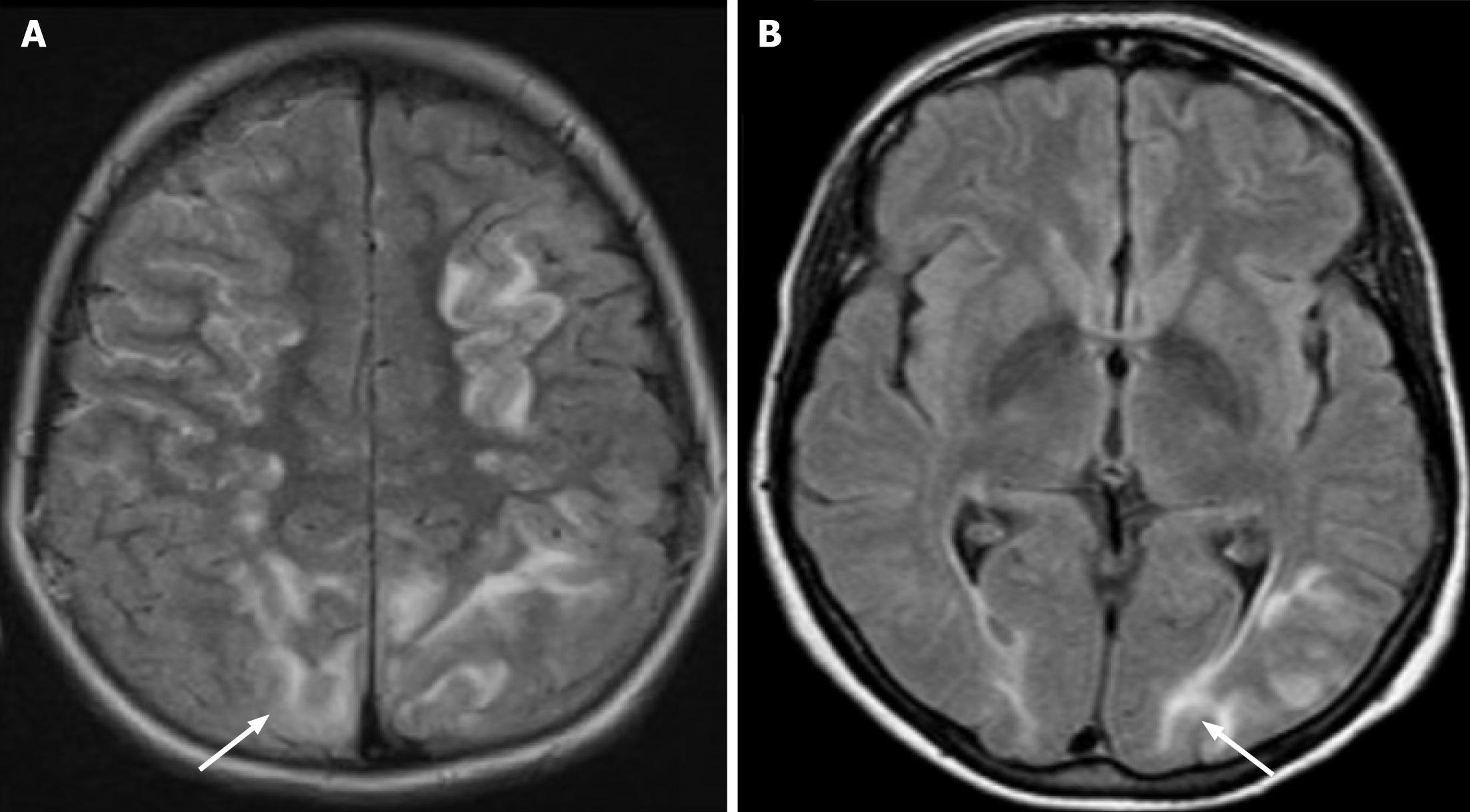
Figure 1 Magnetic resonance imaging of the brain (Axial Fluid-Attenuated Inversion Recovery sequence imaging) showing bilateral cortical and subcortical hyperintense lesions (arrows) involving occipital lobes and parietal lobes.
A: Hyperintense lesions in the parietooccipital sulcus (white arrow); B: Hyperintense lesions at the transverse occipital fasciculi (white arrow).
- Citation: Dilibe A, Subramanian L, Poyser TA, Oriaifo O, Brady R, Srikanth S, Adabale O, Bolaji OA, Ali H. Tacrolimus-induced posterior reversible encephalopathy syndrome following liver transplantation. World J Transplant 2024; 14(2): 91146
- URL: https://www.wjgnet.com/2220-3230/full/v14/i2/91146.htm
- DOI: https://dx.doi.org/10.5500/wjt.v14.i2.91146


