Copyright
©The Author(s) 2025.
World J Rheumatol. Jan 7, 2025; 12(1): 101278
Published online Jan 7, 2025. doi: 10.5499/wjr.v12.i1.101278
Published online Jan 7, 2025. doi: 10.5499/wjr.v12.i1.101278
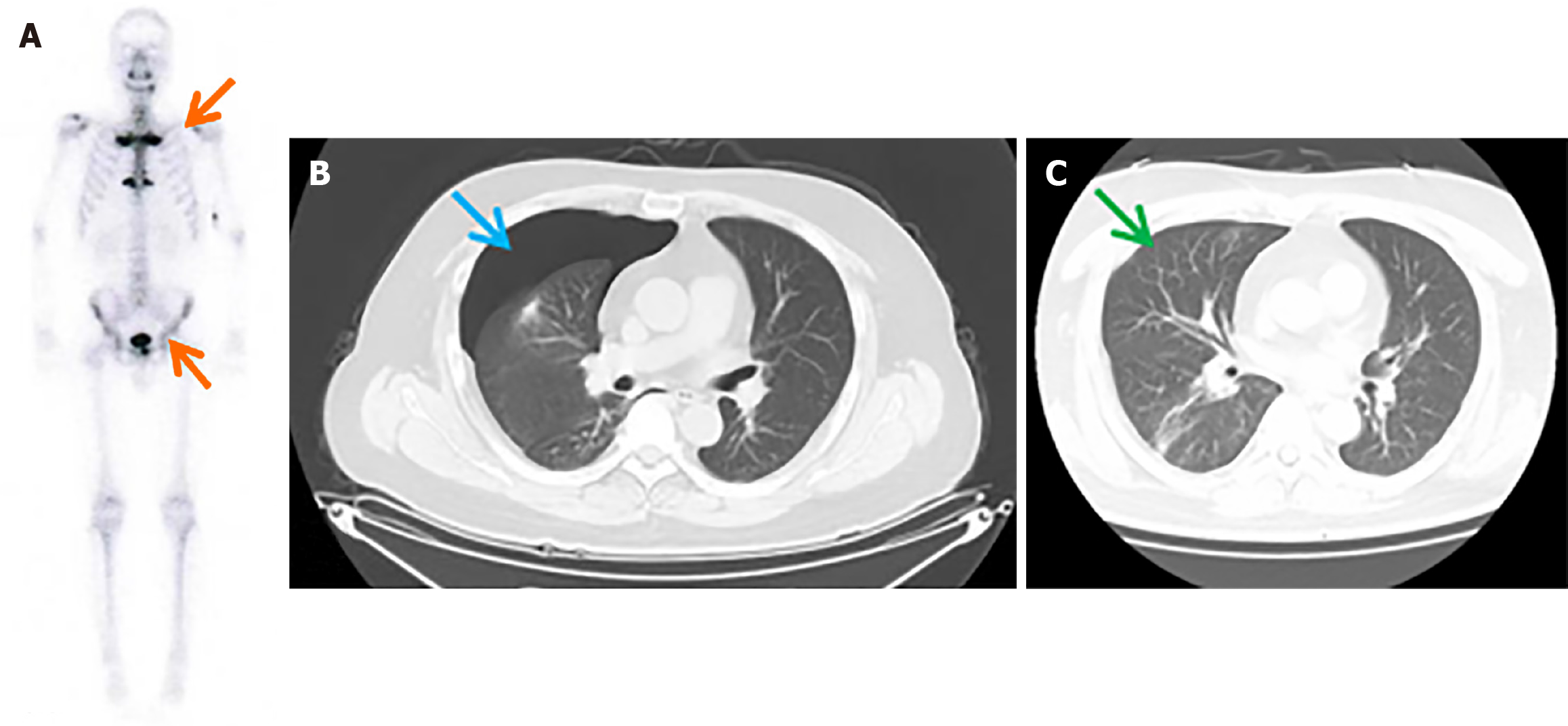
Figure 1 99mTc-MDP bone scintigraphy and pneumothorax images in a patient with synovitis, acne, pustulosis, hyperostosis, and osteitis syndrome.
A: 99mTc-MDP bone scintigraphy revealing abnormal radioactivity accumulation in the anterior chest wall and bilateral sternoclavicular joints (indicated by the orange arrows); B: Computed tomography scan showing a right-sided pneumothorax (indicated by the blue arrow); C: Follow-up chest radiograph, taken one week after completion of pneumothorax treatment, showing resolution (indicated by the green arrow).
- Citation: Zheng ZX, Gu MJ, Kang TL, Zhang YR, Wang YN, Li C, Wu YH. Synovitis, acne, pustulosis, hyperostosis, and osteitis syndrome as a cause of pneumothorax: A case report. World J Rheumatol 2025; 12(1): 101278
- URL: https://www.wjgnet.com/2220-3214/full/v12/i1/101278.htm
- DOI: https://dx.doi.org/10.5499/wjr.v12.i1.101278


