Copyright
©The Author(s) 2022.
World J Psychiatry. May 19, 2022; 12(5): 708-721
Published online May 19, 2022. doi: 10.5498/wjp.v12.i5.708
Published online May 19, 2022. doi: 10.5498/wjp.v12.i5.708
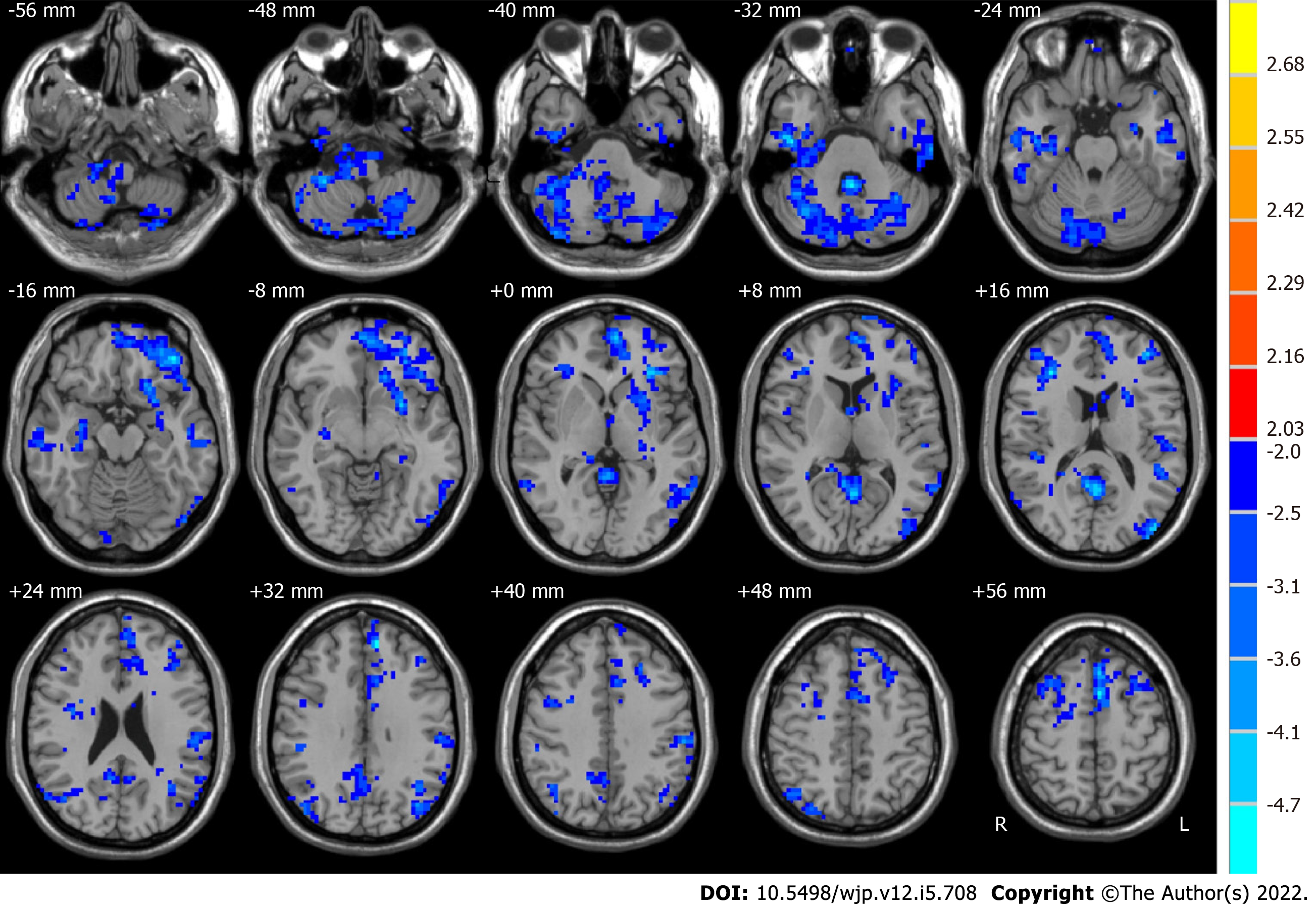
Figure 1 Brain regions with significant alterations in amplitude of low-frequency fluctuations in the typical band (0.
01-0.08 Hz) between healthy controls and pre-electroconvulsive therapy patients. The red region indicates that the amplitude of low-frequency fluctuations in pre-electroconvulsive therapy (ECT) patients was larger than that in healthy controls (HCs). In contrast, the blue region represents HCs that were larger than pre-ECT patients.
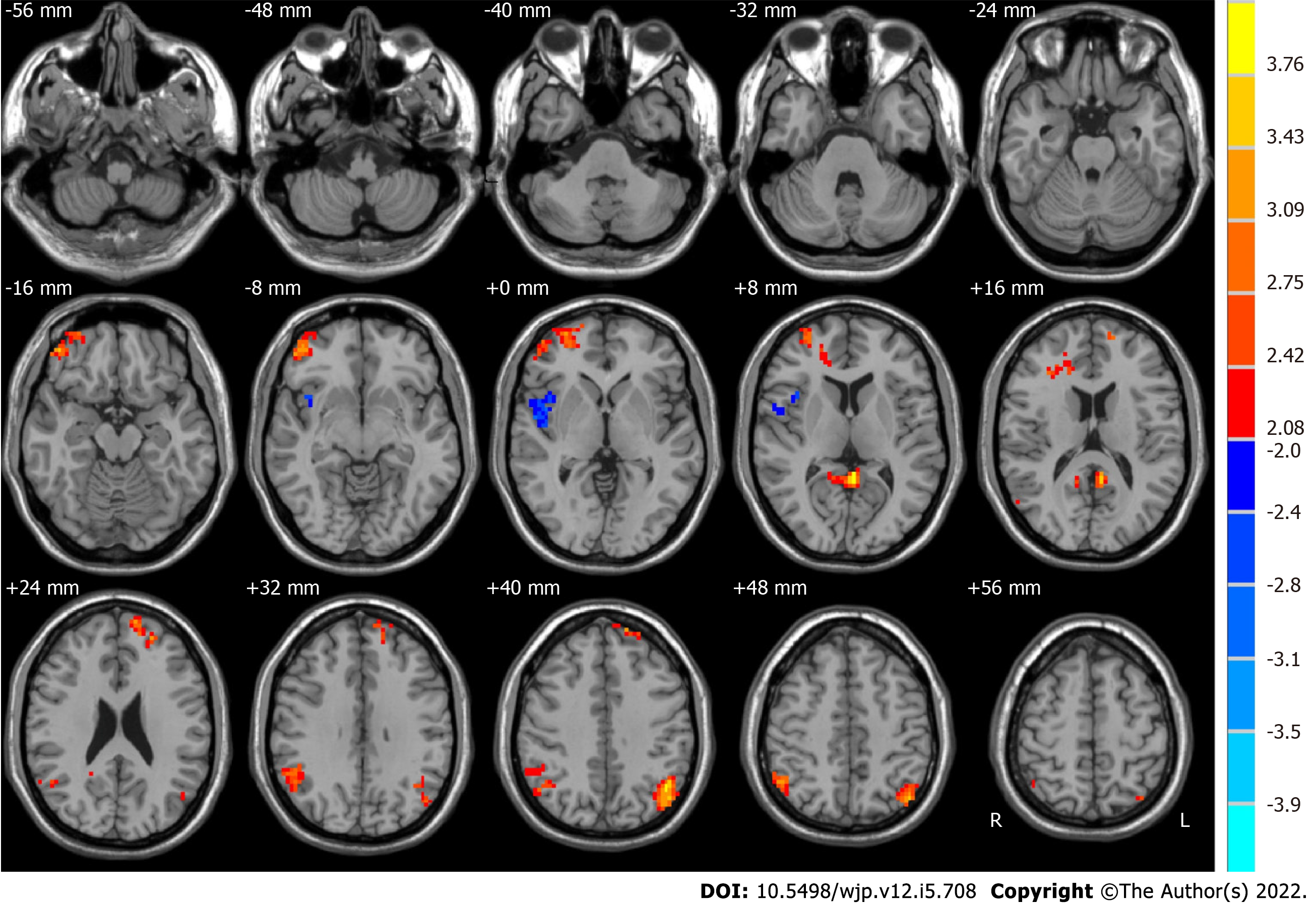
Figure 2 Brain regions with significant alterations in amplitude of low-frequency fluctuations in the typical band (0.
01-0.08 Hz) for pre- and post-electroconvulsive therapy. The red region indicates that the amplitude of low-frequency fluctuations (ALFF) in post-electroconvulsive therapy (ECT) patients was larger than that in pre-ECT patients. In contrast, the blue region represents areas for which the ALFF in pre-ECT patients was larger than that in post-ECT patients.
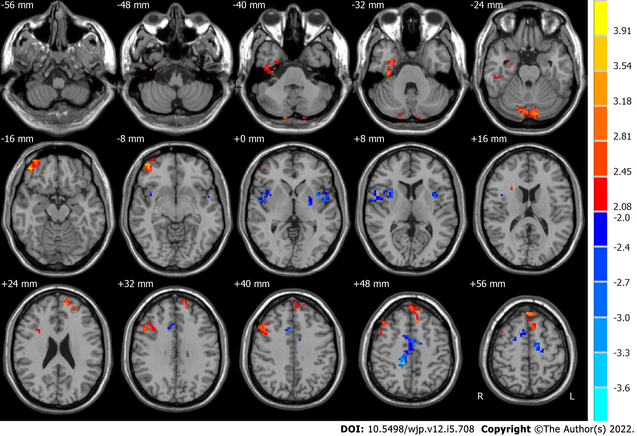
Figure 3 Brain regions with significant alterations in amplitude of low-frequency fluctuations at slow-5 (0.
01–0.027 Hz) for pre- and post-electroconvulsive therapy. The red region indicates that the amplitude of low-frequency fluctuations in post-electroconvulsive therapy (ECT) patients was larger than that in pre-ECT patients. In contrast, the blue region represents pre-ECT patients that were larger than that in post-ECT patients.
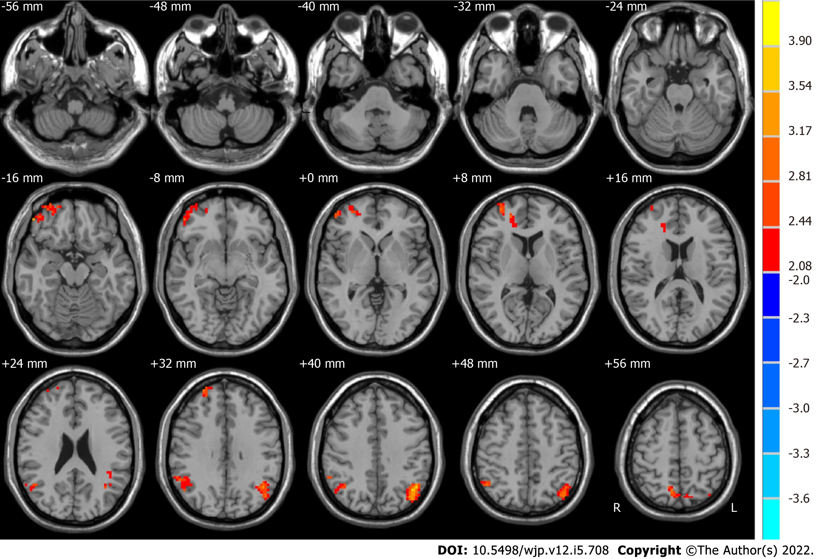
Figure 4 Brain regions with significant alterations in amplitude of low-frequency fluctuations at slow-4 (0.
027-0.08 Hz) for pre- and post-electroconvulsive therapy. The red region indicates that the amplitude of low-frequency fluctuations in post-electroconvulsive therapy (ECT) patients was larger than that in pre-ECT patients. In contrast, the blue region represents pre-ECT patients that were larger than that in post-ECT patients.
- Citation: Li XK, Qiu HT, Hu J, Luo QH. Changes in the amplitude of low-frequency fluctuations in specific frequency bands in major depressive disorder after electroconvulsive therapy. World J Psychiatry 2022; 12(5): 708-721
- URL: https://www.wjgnet.com/2220-3206/full/v12/i5/708.htm
- DOI: https://dx.doi.org/10.5498/wjp.v12.i5.708


