Copyright
©The Author(s) 2016.
World J Hypertens. May 23, 2016; 6(2): 66-75
Published online May 23, 2016. doi: 10.5494/wjh.v6.i2.66
Published online May 23, 2016. doi: 10.5494/wjh.v6.i2.66
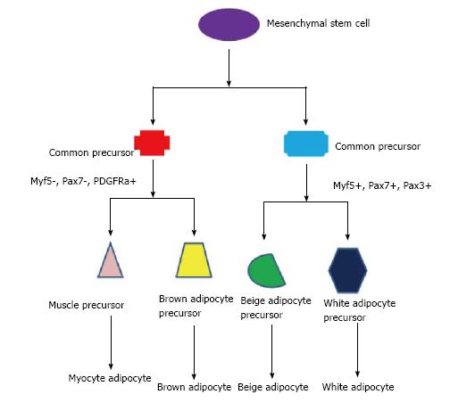
Figure 1 Schematic demonstrating links between three basic type of adipocytes and muscle.
All cells come from the same common cell, mesenchymal stem cell. Presence or absence of certain factors during development, such as Myf5, Pax 3 and 7, PDGFRa and myogenin can affect the final product of the cell. Myf5: Myogenic factor 5; Pax 7: Paired box protein 7; Pax 3: Paired box protein 3; PDGFRa: Platelet-derived growth factor receptor, alpha polypeptide.
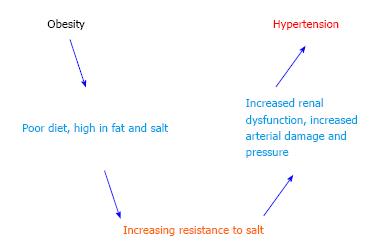
Figure 2 Schematic demonstrating potential relationship between obesity and hypertension.
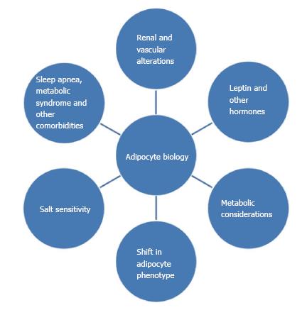
Figure 3 Visual representation of factors contributing to the complex pathophysiology linking adipocyte biology and hypertension.
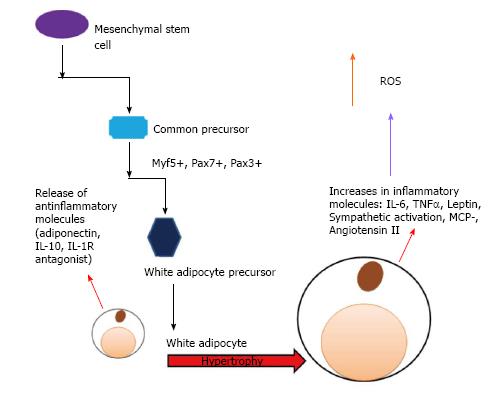
Figure 4 Small adipocytes release anti-inflammatory agents such as adiponectin, interleukin 10, and interleukin 1 receptor antagonist.
Hypertrophy of small adipocytes into large adipocytes changes the biochemical release products, into inflammatory markers [interleukin-6 (IL-6), tumor necrosis factor alpha (TNFα), and monocyte chemotactic protein 1 (MCP1)] and other chemicals such as leptin and angiotensin II that contribute to the disease. Increased activation of the sympathetic nervous system also contributes. IL-10: Interleukin-10; IL-1R: Interleukin 1 receptor; Myf5: Myogenic factor 5; Pax 7: Paired box protein 7; Pax 3: Paired box protein 3; ROS: Reactive oxygen species.
- Citation: Martin R, Shapiro JI. Role of adipocytes in hypertension. World J Hypertens 2016; 6(2): 66-75
- URL: https://www.wjgnet.com/2220-3168/full/v6/i2/66.htm
- DOI: https://dx.doi.org/10.5494/wjh.v6.i2.66


