Minireviews Open Access
Copyright ©The Author(s) 2024. Published by Baishideng Publishing Group Inc. All rights reserved.
World J Exp Med. Sep 20, 2024; 14(3): 97233
Published online Sep 20, 2024. doi: 10.5493/wjem.v14.i3.97233
Anti-aging based on stem cell therapy: A scoping review
Nassar El Assaad, Nour Bou Atme, Samer Abou Zeid, Marwan Ghosn, Faculty of Medicine, Saint Joseph University, Beirut 961, Lebanon
Alain Chebly, Centre Jacques Loiselet for Medical Genetics and Genomics (CGGM), Faculty of Medicine, Saint Joseph University, Beirut 961, Lebanon
Alain Chebly, Higher Institute of Public Health, Saint Joseph University, Beirut 961, Lebanon
Rawad Salame, Khalil Akouch, Charbel Khalil, Stem Cell Therapy Lab, Reviva Regenerative Medicine Center, Beirut 961, Lebanon
Robert Achkar, Poz Pozan University of Medical Sciences, Pozan 034, Poland
Paul Rafoul, Department of Epidemiology and Biostatistics, Faculty of Public Health, Lebanese University, Beirut 961, Lebanon
Colette Hanna, Charbel Khalil, School of Medicine, Lebanese American University, Beirut 961, Lebanon
Charbel Khalil, Bone Marrow Transplant Unit, Burjeel Medical City, Abu Dhabi 999041, United Arab Emirates
ORCID number: Charbel Khalil (0000-0001-7740-6963).
Author contributions: Khalil C was responsible for conceptualization and supervision; Ghosn M, Khalil C, Chebly A and Bou Atme N were responsible for methodology; Khalil C and Rafoul P were responsible for validation; El Assaad N and Khalil Cwere responsible for formal analysis, data curation; Khalil C and Akouch K were responsible for writing-original draft preparation; Akouch K, Achkar R, Bou Atme N and Khalil Cwere responsible for visualization; El Assaad N, Hanna C, Salame R, Ghosn M, Abou Zeid S, Achkar R, Raffoul P, Hanna C, Chebly A and Khalil C were responsible for writing - review & editing; all authors have read and agreed to the submitted version of the manuscript.
Conflict-of-interest statement: The authors declare that they have no conflict of interest.
Open-Access: This article is an open-access article that was selected by an in-house editor and fully peer-reviewed by external reviewers. It is distributed in accordance with the Creative Commons Attribution NonCommercial (CC BY-NC 4.0) license, which permits others to distribute, remix, adapt, build upon this work non-commercially, and license their derivative works on different terms, provided the original work is properly cited and the use is non-commercial. See: https://creativecommons.org/Licenses/by-nc/4.0/
Corresponding author: Charbel Khalil, PhD, Director, Stem Cell Therapy Lab, Reviva Regenerative Medicine Center, Bsalim, Main Road Lebanon, Beirut 961, Beyrouth, Lebanon. c.khalil@revivamedical.net
Received: May 26, 2024
Revised: July 4, 2024
Accepted: July 18, 2024
Published online: September 20, 2024
Processing time: 94 Days and 13.9 Hours

Abstract

Stem cells are present in the tissues and organs and remain in a quiescent and undifferentiated state until it is physiologically necessary to produce new descendant cells. Due to their multipotency property, mesenchymal stem cells have attracted considerable attention worldwide due to their immunomodulation and therapeutic function in tissue regeneration. Stem cells secrete components such as paracrine factors, extracellular vesicles, and exosomes which have been shown to have anti-inflammatory, anti-aging, reconstruction and wound healing potentials in many in vitro and in vivo models. The pluripotency and immunomodulatory features of stem cells could potentially be an effective tool in cell therapy and tissue repair. Aging affects the capacity for self-renewal and differentiation of stem cells, decreasing the potential for regeneration and the loss of optimal functions in organisms over time. Current progress in the field of cellular therapy and regenerative medicine has facilitated the evolution of particular guidelines and quality control approaches, which eventually lead to clinical trials. Cell therapy could potentially be one of the most promising therapies to control aging due to the fact that single stem cell transplantation can regenerate or substitute the injured tissue. To understand the involvement of stem cells not only in tissue maintenance and disease but also in the control of aging it is important to know and identify their properties, functions, and regulation in vivo, which are addressed in this review.

Key Words: Mesenchymal stem cell; Anti-aging; Telomerase; Regenerative medicine; Stem cell therapy

Core Tip: Mesenchymal stem cells (MSCs) exhibit promising anti-aging properties by targeting the underlying mechanisms of aging, including chronic inflammation, cellular senescence, and oxidative stress. Through their regenerative potential, immunomodulatory abilities, and secretome production, MSCs contribute to tissue repair and rejuvenation. These unique capabilities position MSCs as key players in the future of anti-aging therapies and regenerative medicine, offering potential interventions for promoting healthier and more vibrant aging.


INTRODUCTION

Aging is the time-related deterioration of essential physiological functions for survival and fertility, occurring universally in various organisms and leading to natural death. This process involves gradual dysfunctions in all organs, affecting unicellular organisms, plants, animals, and humans alike. It is an inevitable stage of development and life. Zhang et al[1] define aging as the reduction of the body's capacity, both physically and psychologically, to adapt to the environment, and the gradual tendency towards death.

Recent gerontological research has delved into the complex biochemical mechanisms associated with the gradual decline in bodily functions. It emphasizes the involvement of multiple processes at the cellular, molecular, and systemic level, requiring comprehensive approaches for understanding aging. Despite being a physiological and universal process, aging occurs at an individual rate in each person[2,3].

Differentiating between chronological and biological age is important due to the varying rates of aging. Chronological age merely indicates the time passed since birth, while biological age encompasses a broad spectrum of physical, physiological, and cognitive functions, influenced by molecular and cellular processes[4]. Despite ongoing discussions, the biology of aging remains controversial, posing challenges in establishing a universally accepted definition of normal aging[5]. However, biological aging relies on various theories, including those related to the effects of free radicals, telomere shortening, and the mitochondrial theory.

In the broader context of anti-aging research, healthy aging is characterized by the attainment of an extended period of robust physiological and mental well-being. This entails an ongoing effort to seize opportunities for enhancing physical and mental health, preserving independence, and elevating overall quality of life across the lifespan. Seals et al[6] proposed the concept of optimal longevity, incorporating a compressed disease period at life's end. This innovative perspective has led to the emergence of geroscience, a new field in aging research. Geroscience is dedicated to identifying and intervening in biological mechanisms to enhance health span and actively promote healthy aging in individuals.

Aging itself is initiated by a combination of genetic and environmental factors that can influence organisms from birth. The signs of aging encompass various aspects, including impaired vision, hearing loss, muscle strength decline, reduced bone density, weakened immune system, cognitive decline, less efficient metabolism, reduced energy, hair loss, diminished balance, and overall decreased mobility[7].

Understanding these aging indicators is crucial within the setting of geroscience in order to promote extended health span and well-being throughout the aging process. In this review, we analyze the multifaceted roles of stem cells in tissue maintenance, disease pathogenesis, and the regulation of aging by comprehensively examining their properties, functions, and regulatory mechanisms in vivo.

AGING AND IMPAIRED VISION

Older adults commonly face three visual problems: Impaired spatial contrast sensitivity, scotopic sensitivity loss, and delayed rod-mediated dark adaptation, along with reduced visual processing speed. While the extent of deficits varies among individuals, older adults are likely to experience one or more of these disturbances. These visual challenges impact daily tasks. In severe cases, these aging-related visual issues may indicate the emergence of visual pathway conditions and diseases common in the elderly[8].

AGING AND IMPAIRED HEARING

Age-related hearing loss (ARHL), or presbycusis, is a common occurrence in mammals, including humans, with varying onset times and degrees of loss. It manifests through reduced sensitivity to sound, especially high pitches, and a diminished ability to understand speech in background noise[9].

ARHL involves both peripheral structures of the inner ear and central acoustic pathways, with oxidative stress identified as a key pro-aging mechanism in the human cochlea[10].

LOSS OF BONE DENSITY AND MUSCLE STRENGTH

Muscle and bone aging contribute significantly to morbidity and mortality in older populations, affecting the overall quality of life. Skeletal aging begins after reaching peak bone mass, with varying onset ages and rates between sexes. Two primary factors contribute to the decline in muscle mass and function with age: Muscle fiber atrophy and muscle fiber loss. It is evident that both these components exert influence on the regulation of muscle atrophy and dysfunction, impacting either individual muscles or groups of muscles[11].

Bone loss is accelerated during the perimenopausal period in women and gradually progresses in men of advanced age. Changes in both bone quantity and quality occur throughout growth and aging, impacting microarchitecture, size, and geometry. Genetic and epigenetic factors may predispose individuals to osteoporosis, characterized by weakened bones and an increased risk of fractures[12].

WEAKENED IMMUNE SYSTEM

The concept of immune senescence reflects age-related changes in immune responses, both cellular and serological, affecting the process of generating specific responses to foreign and self-antigens. The decline of the immune system with age is reflected in the increased susceptibility to infectious diseases, weaker responses to vaccination, increased prevalence of cancer, autoimmune and other chronic diseases[13].

AGING AT THE CELLULAR LEVEL

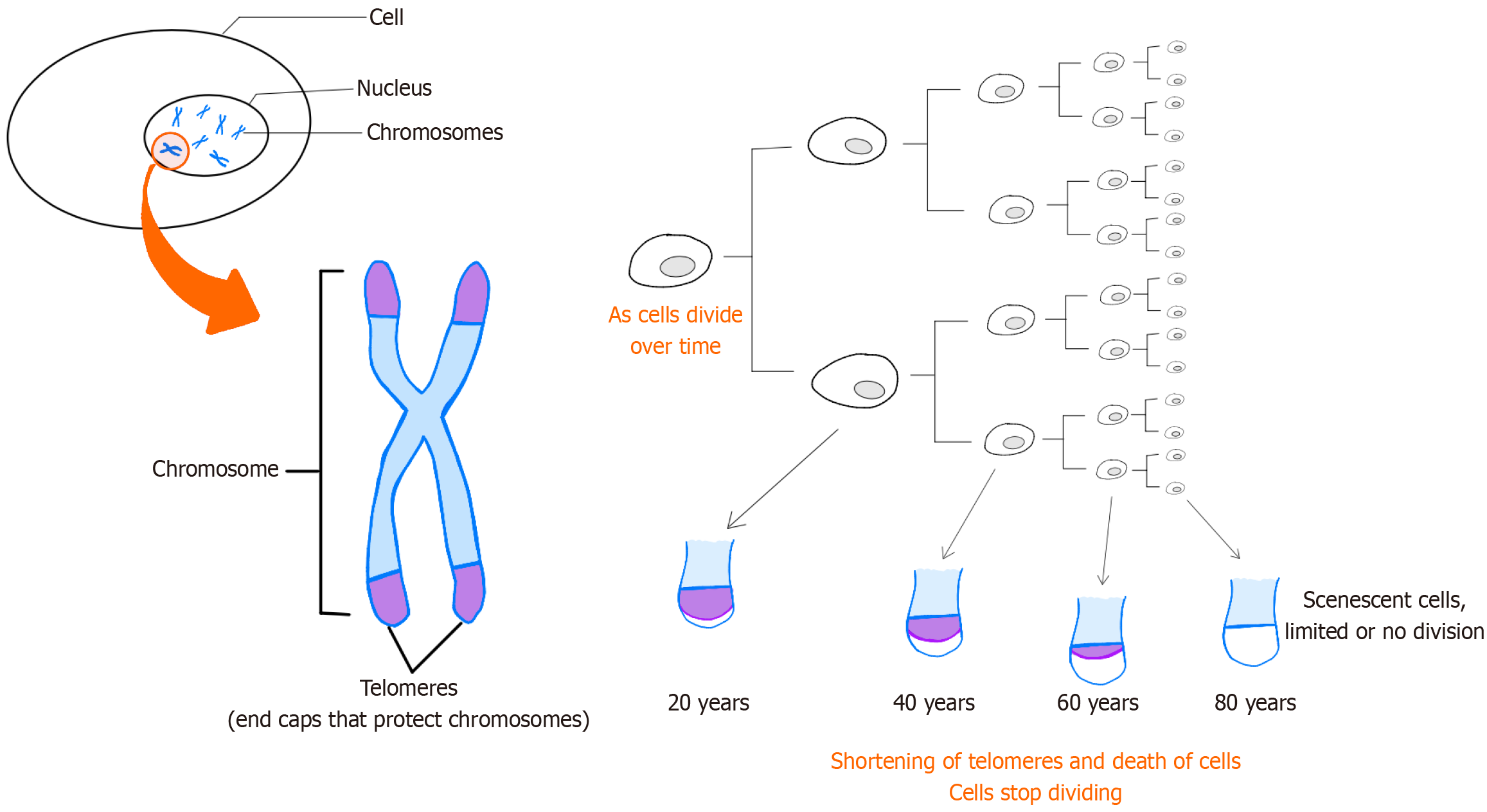
In 2013, López-Otín et al[14] defined nine cellular and molecular hallmarks of aging, laying a crucial foundation for future research in the field. These hallmarks encompass genomic instability, telomere attrition, epigenetic alterations, loss of proteostasis, deregulated nutrient-sensing, mitochondrial dysfunction, cellular senescence, stem cell exhaustion, and altered intercellular communication (Figure 1). Expanding on this framework, a research symposium entitled "New Hallmarks of Aging" convened in Copenhagen on March 22, 2022. This symposium concentrated on presenting innovative findings and recontextualizing the original nine hallmarks, introducing potential new hallmarks such as compromised autophagy, dysregulation of RNA processing, microbiome disturbances, altered mechanical properties, and inflammation[14,15].

Figure 1
Figure 1 Telomeres on the end of chromosomes are believed to protect the DNA strands and prevent them from fusing with other strands. Telomeres lose a little of their length during each cell division. Since replicative DNA polymerases are not able to replicate telomeres, and telomerase is not expressed in normal mammalian somatic cells, telomeres become too short to replicate after a fixed number of cell divisions. Eventually, the cell will stop growing and enter cellular senescence.
STEM CELLS AND MESENCHYMAL STEM CELLS IN AGING

Stem cells, characterized by their immature nature, possess the remarkable ability to undergo infinite self-renewal and differentiate into various cell lineages. Their unique capacity to function as a reservoir for the production, maintenance, repair, and regeneration of diverse tissues distinguishes them from other cell types[16].

There are two main subtypes of stem cells: Embryonic stem cells, derived from early-stage embryos, and mesenchymal stem cells (MSCs), found in adults and isolated from various tissues such as bone marrow and adipose tissue.

As a subtype of adult stem cells, MSCs are multipotent stem cells with remarkable regenerative potential found in various tissues throughout the body. They play a pivotal role in tissue regeneration and can differentiate into multiple cell lineages. Amongst the myriad roles played by MSCs, one of the most captivating and promising aspects lies in their remarkable ability to counteract the effects of aging. MSCs can go beyond their traditional roles in tissue repair, immune modulation, and paracrine signaling to intricately engage in fighting the multifaceted processes associated with aging, making them particularly promising for future clinical trials, especially in the fields of regenerative medicine and anti-aging treatments[17-19].

In fact, MSCs are known for their important proliferation, especially if the donor is young since proliferation capacity declines with age. Also, they can be differentiated into osteoblasts, chondrocytes, myoblasts, adipocytes, and fibroblasts. Additionally, MSCs exhibited anti-aging benefits through the secretion of cytokines and growth factors that act as promoters of angiogenesis, anti-inflammatory agents, and inhibitors of apoptosis[20,21].

In a recent study, Wang et al[22] highlighted the anti-aging and anti-obesity effects of MSCs from healthy mice when injected into older mice, providing new insights into potential anti-aging treatments. Moreover, MSCs, especially adipose stem cells, have been reported to improve aged skin by increasing angiogenesis growth factors[23].

MSCs reportedly release a wide array of bioactive molecules that collectively form the secretome. Within this complex mixture, an important subset is the exosomes which are tiny vesicles enriched with molecular cargo. These extracellular components, derived from the endosomal pathway within MSCs, constitute a vital facet of the broader secretome. The secretome, encompassing soluble factors such as growth factors and cytokines alongside exosomes, acts as a comprehensive communication system. Through paracrine signaling, MSCs influence neighboring and distant cells, fostering an environment conducive to tissue repair, immunomodulation, and anti-inflammatory responses. The interplay between MSCs, secretome, and exosomes highlights the interconnected web of regenerative mechanisms, underscoring their potential in therapeutic applications, from promoting wound healing to addressing aging-related degeneration[24-28].

These anti-aging effects encompass a spectrum of activities, from mitigating chronic inflammation to enhancing tissue regeneration and modulating cellular senescence[29]. The unique capacity of MSCs to address the underlying mechanisms of aging positions them at the forefront of regenerative medicine, holding immense potential for interventions, aiming to promote not just longevity, but a healthier and more vibrant aging process[30,31].

TISSUE REGENERATION AND STEM CELL DIFFERENTIATION

MSCs are a subset of non-hematopoietic adult stem cells capable of differentiating into various cell types, including osteoblasts, chondrocytes, and adipocytes. This differentiation capacity and immunomodulatory properties contribute to tissue regeneration by replacing damaged or aging cells with new, functional cells. The differentiation potential of MSCs makes them promising candidates for regenerative medicine applications in repairing fragile tissues associated with the musculoskeletal system, nervous system, myocardium, liver, cornea, trachea, and skin[32-34].

SECRETOME PRODUCTION AND IMMUNOMODULATION

As chronic inflammation is a hallmark of aging, MSCs exhibit immunomodulatory properties, suppressing excessive inflammatory responses by release of the secretome which contains a variety of anti-inflammatory cytokines and growth factors. These factors stimulate cell proliferation, enhance tissue repair, and contribute to overall health by modulating the immune system and creating an anti-inflammatory environment[35-37].

Intercellular communication

Exosomes, derived from MSCs, contain bioactive molecules, including proteins, lipids, and nucleic acids. They act as messengers, transferring information between cells which can influence neighboring cells, promoting tissue repair and rejuvenation[38,39].

Anti-fibrotic effects

MSCs and their secretome may have anti-fibrotic effects, reducing the accumulation of fibrotic tissue in organs. Fibrosis is associated with aging leading to impaired organ function. Hence, the therapeutic potential of MSCs resides in their capability to target multiple fibrogenesis parameters. This includes their capacity for immunosuppression, inhibition of the TGF-β1 pathway and mitigation of hypoxia and oxidative stress[40].

Cellular protection

MSC-derived exosomes may carry antioxidant enzymes and other protective factors. This cargo can help protect cells from oxidative stress, which is associated with cellular damage and an accelerated aging process[41].

Mitochondrial function

MSCs and exosomes may influence mitochondrial function, by enhancing energy production within cells. Indeed, improved mitochondrial function is associated with increased cellular resilience and longevity[42,43].

Senescence modulation

MSCs and exosomes may modulate cellular senescence, the process by which cells lose their ability to divide and function properly. By influencing senescence, MSCs contribute to maintaining youthful cellular functions[36,44].

Extracellular matrix remodeling

MSCs and secretome contribute to remodeling of the extracellular matrix, ensuring the maintenance of tissue structure, elasticity, and functionality[45].

Stem cell niche maintenance

MSCs and secretome play a role in maintaining stem cell niches in tissues. This ensures a continuous pool of functional cells for tissue repair and regeneration[46].

Enhanced wound healing

MSCs and exosomes contribute to enhanced wound healing, which is essential for preventing age-related complications and maintaining the integrity of the skin and other tissues in the elderly[38,46].

Angiogenesis promotion

Growth factors found in the secretome can stimulate angiogenesis and the formation of new blood vessels. Improved blood flow is crucial for supplying nutrients to tissues and therefore supporting overall healthy tissues[47,48].

Promotion of autophagy

Exosomes may stimulate autophagy, a cellular process that removes damaged components. Enhanced autophagy can contribute to cellular rejuvenation, thereby underlining their therapeutic efficacy. Consequently, strategies aimed at modulating autophagy through MSC-based therapies hold significant promise for enhancing therapeutic outcomes in various human diseases, including cancer, autoimmune disorders, and neurodegenerative conditions[49,50].

CONCLUSION

As we delve into the intricacies of MSCs and their multifaceted roles, it becomes evident that these cells hold immense promise in revolutionizing the landscape of regenerative medicine especially in the elderly. MSCs have important characteristics that make them ideal candidates for use in regenerative medicine, such as immunomodulatory capability valuable for improving immune system abnormalities, paracrine or autocrine roles that produce growth factors, and the vital potential to differentiate into various cells. Also, there is a lack of anti-aging treatment derived from the mechanism of aging. This review showed that MSCs have a significant effect on delaying aging. This exploration of their roles provides a foundation for understanding their potential applications in addressing a wide array of health-related diseases and challenges. It is essential to note that research in this field is ongoing, and clinical applications are still being explored. Further studies are needed to understand the optimal conditions for MSC-based therapies and their long-term effects on aging-related conditions.

ACKNOWLEDGEMENTS

We thank all who contributed to this work from all institutions.

Footnotes

Provenance and peer review: Unsolicited article; Externally peer reviewed.

Peer-review model: Single blind

Specialty type: Medicine, research and experimental

Country of origin: Lebanon

Peer-review report’s classification

Scientific Quality: Grade C

Novelty: Grade B

Creativity or Innovation: Grade C

Scientific Significance: Grade C

P-Reviewer: Bonartsev A S-Editor: Lin C L-Editor: Webster JR P-Editor: Wang WB

References
1.  Zhang Y, Li Q, Niu Y, Wei K, Wang X, Niu B, Zhang L. Research progress on aging mechanism and drugs and the role of stem cells in anti-aging process. Exp Gerontol. 2023;179:112248.  [PubMed]  [DOI]  [Cited in This Article: ]  [Cited by in F6Publishing: 2]  [Reference Citation Analysis (0)]
2.  Park DC, Yeo SG. Aging. Korean J Audiol. 2013;17:39-44.  [PubMed]  [DOI]  [Cited in This Article: ]  [Cited by in Crossref: 34]  [Cited by in F6Publishing: 32]  [Article Influence: 2.9]  [Reference Citation Analysis (0)]
3.  Hayflick L. Biological aging is no longer an unsolved problem. Ann N Y Acad Sci. 2007;1100:1-13.  [PubMed]  [DOI]  [Cited in This Article: ]  [Cited by in Crossref: 255]  [Cited by in F6Publishing: 223]  [Article Influence: 13.1]  [Reference Citation Analysis (0)]
4.  Ferrucci L, Gonzalez-Freire M, Fabbri E, Simonsick E, Tanaka T, Moore Z, Salimi S, Sierra F, de Cabo R. Measuring biological aging in humans: A quest. Aging Cell. 2020;19:e13080.  [PubMed]  [DOI]  [Cited in This Article: ]  [Cited by in Crossref: 392]  [Cited by in F6Publishing: 323]  [Article Influence: 80.8]  [Reference Citation Analysis (0)]
5.  Cohen AA, Legault V, Fülöp T. What if there's no such thing as "aging"? Mech Ageing Dev. 2020;192:111344.  [PubMed]  [DOI]  [Cited in This Article: ]  [Cited by in Crossref: 16]  [Cited by in F6Publishing: 17]  [Article Influence: 4.3]  [Reference Citation Analysis (0)]
6.  Seals DR, Justice JN, LaRocca TJ. Physiological geroscience: targeting function to increase healthspan and achieve optimal longevity. J Physiol. 2016;594:2001-2024.  [PubMed]  [DOI]  [Cited in This Article: ]  [Cited by in Crossref: 144]  [Cited by in F6Publishing: 175]  [Article Influence: 19.4]  [Reference Citation Analysis (0)]
7.  Amarya S, Singh K, Sabharwal M. Ageing Process and Physiological Changes. Gerontology. 2018;.  [PubMed]  [DOI]  [Cited in This Article: ]  [Cited by in Crossref: 26]  [Cited by in F6Publishing: 27]  [Article Influence: 4.5]  [Reference Citation Analysis (0)]
8.  Owsley C. Aging and vision. Vision Res. 2011;51:1610-1622.  [PubMed]  [DOI]  [Cited in This Article: ]  [Cited by in Crossref: 411]  [Cited by in F6Publishing: 356]  [Article Influence: 27.4]  [Reference Citation Analysis (0)]
9.  Bowl MR, Dawson SJ. Age-Related Hearing Loss. Cold Spring Harb Perspect Med. 2019;9.  [PubMed]  [DOI]  [Cited in This Article: ]  [Cited by in Crossref: 185]  [Cited by in F6Publishing: 164]  [Article Influence: 32.8]  [Reference Citation Analysis (0)]
10.  Guerrieri M, Di Mauro R, Di Girolamo S, Di Stadio A. Hearing and Ageing. Subcell Biochem. 2023;103:279-290.  [PubMed]  [DOI]  [Cited in This Article: ]  [Reference Citation Analysis (0)]
11.  Wilkinson DJ, Piasecki M, Atherton PJ. The age-related loss of skeletal muscle mass and function: Measurement and physiology of muscle fibre atrophy and muscle fibre loss in humans. Ageing Res Rev. 2018;47:123-132.  [PubMed]  [DOI]  [Cited in This Article: ]  [Cited by in Crossref: 216]  [Cited by in F6Publishing: 357]  [Article Influence: 59.5]  [Reference Citation Analysis (0)]
12.  Goltzman D  The Aging Skeleton. In: Rhim J, Dritschilo A, Kremer R, editors. Human Cell Transformation. Cham: Springer International Publishing, 2019: 153-160.  [PubMed]  [DOI]  [Cited in This Article: ]
13.  Castelo-Branco C, Soveral I. The immune system and aging: a review. Gynecol Endocrinol. 2014;30:16-22.  [PubMed]  [DOI]  [Cited in This Article: ]  [Cited by in Crossref: 249]  [Cited by in F6Publishing: 245]  [Article Influence: 24.5]  [Reference Citation Analysis (0)]
14.  López-Otín C, Blasco MA, Partridge L, Serrano M, Kroemer G. The hallmarks of aging. Cell. 2013;153:1194-1217.  [PubMed]  [DOI]  [Cited in This Article: ]  [Cited by in Crossref: 10570]  [Cited by in F6Publishing: 9251]  [Article Influence: 841.0]  [Reference Citation Analysis (0)]
15.  Chebly A, Khalil C, Kuzyk A, Beylot-Barry M, Chevret E. T-cell lymphocytes' aging clock: telomeres, telomerase and aging. Biogerontology. 2024;25:279-288.  [PubMed]  [DOI]  [Cited in This Article: ]  [Cited by in Crossref: 1]  [Reference Citation Analysis (0)]
16.  Schmauck-Medina T, Molière A, Lautrup S, Zhang J, Chlopicki S, Madsen HB, Cao S, Soendenbroe C, Mansell E, Vestergaard MB, Li Z, Shiloh Y, Opresko PL, Egly JM, Kirkwood T, Verdin E, Bohr VA, Cox LS, Stevnsner T, Rasmussen LJ, Fang EF. New hallmarks of ageing: a 2022 Copenhagen ageing meeting summary. Aging (Albany NY). 2022;14:6829-6839.  [PubMed]  [DOI]  [Cited in This Article: ]  [Cited by in Crossref: 1]  [Cited by in F6Publishing: 127]  [Article Influence: 63.5]  [Reference Citation Analysis (0)]
17.  Laplane L, Solary E. Towards a classification of stem cells. Elife. 2019;8.  [PubMed]  [DOI]  [Cited in This Article: ]  [Cited by in Crossref: 16]  [Cited by in F6Publishing: 23]  [Article Influence: 4.6]  [Reference Citation Analysis (0)]
18.  Glenn JD, Whartenby KA. Mesenchymal stem cells: Emerging mechanisms of immunomodulation and therapy. World J Stem Cells. 2014;6:526-539.  [PubMed]  [DOI]  [Cited in This Article: ]  [Cited by in Crossref: 293]  [Cited by in F6Publishing: 279]  [Article Influence: 27.9]  [Reference Citation Analysis (1)]
19.  Parekkadan B, Milwid JM. Mesenchymal stem cells as therapeutics. Annu Rev Biomed Eng. 2010;12:87-117.  [PubMed]  [DOI]  [Cited in This Article: ]  [Cited by in Crossref: 533]  [Cited by in F6Publishing: 556]  [Article Influence: 39.7]  [Reference Citation Analysis (0)]
20.  Margiana R, Markov A, Zekiy AO, Hamza MU, Al-Dabbagh KA, Al-Zubaidi SH, Hameed NM, Ahmad I, Sivaraman R, Kzar HH, Al-Gazally ME, Mustafa YF, Siahmansouri H. Clinical application of mesenchymal stem cell in regenerative medicine: a narrative review. Stem Cell Res Ther. 2022;13:366.  [PubMed]  [DOI]  [Cited in This Article: ]  [Cited by in Crossref: 1]  [Cited by in F6Publishing: 98]  [Article Influence: 49.0]  [Reference Citation Analysis (0)]
21.  Godic A. The role of stem cells in anti-aging medicine. Clin Dermatol. 2019;37:320-325.  [PubMed]  [DOI]  [Cited in This Article: ]  [Cited by in Crossref: 10]  [Cited by in F6Publishing: 10]  [Article Influence: 2.0]  [Reference Citation Analysis (0)]
22.  Wang T, Li Y, Zhu Y, Liu Z, Huang L, Zhao H, Zhou Z, Wu Q. Anti-aging mechanism of different age donor-matched adipose-derived stem cells. Stem Cell Res Ther. 2023;14:192.  [PubMed]  [DOI]  [Cited in This Article: ]  [Reference Citation Analysis (0)]
23.  Ding DC, Shyu WC, Lin SZ. Mesenchymal stem cells. Cell Transplant. 2011;20:5-14.  [PubMed]  [DOI]  [Cited in This Article: ]  [Cited by in Crossref: 352]  [Cited by in F6Publishing: 487]  [Article Influence: 37.5]  [Reference Citation Analysis (0)]
24.  Panda B, Sharma Y, Gupta S, Mohanty S. Mesenchymal Stem Cell-Derived Exosomes as an Emerging Paradigm for Regenerative Therapy and Nano-Medicine: A Comprehensive Review. Life (Basel). 2021;11.  [PubMed]  [DOI]  [Cited in This Article: ]  [Cited by in Crossref: 4]  [Cited by in F6Publishing: 12]  [Article Influence: 4.0]  [Reference Citation Analysis (0)]
25.  Zhang Y, Liu Y, Liu H, Tang WH. Exosomes: biogenesis, biologic function and clinical potential. Cell Biosci. 2019;9:19.  [PubMed]  [DOI]  [Cited in This Article: ]  [Cited by in Crossref: 651]  [Cited by in F6Publishing: 1123]  [Article Influence: 224.6]  [Reference Citation Analysis (0)]
26.  Kandeel M, Morsy MA, Alkhodair KM, Alhojaily S. Mesenchymal Stem Cell-Derived Extracellular Vesicles: An Emerging Diagnostic and Therapeutic Biomolecules for Neurodegenerative Disabilities. Biomolecules. 2023;13.  [PubMed]  [DOI]  [Cited in This Article: ]  [Reference Citation Analysis (0)]
27.  Karnas E, Dudek P, Zuba-Surma EK. Stem cell- derived extracellular vesicles as new tools in regenerative medicine - Immunomodulatory role and future perspectives. Front Immunol. 2023;14:1120175.  [PubMed]  [DOI]  [Cited in This Article: ]  [Cited by in F6Publishing: 11]  [Reference Citation Analysis (0)]
28.  Gurunathan S, Kang MH, Jeyaraj M, Qasim M, Kim JH. Review of the Isolation, Characterization, Biological Function, and Multifarious Therapeutic Approaches of Exosomes. Cells. 2019;8.  [PubMed]  [DOI]  [Cited in This Article: ]  [Cited by in Crossref: 440]  [Cited by in F6Publishing: 668]  [Article Influence: 133.6]  [Reference Citation Analysis (0)]
29.  Li X, Li C, Zhang W, Wang Y, Qian P, Huang H. Inflammation and aging: signaling pathways and intervention therapies. Signal Transduct Target Ther. 2023;8:239.  [PubMed]  [DOI]  [Cited in This Article: ]  [Cited by in Crossref: 27]  [Cited by in F6Publishing: 115]  [Article Influence: 115.0]  [Reference Citation Analysis (0)]
30.  Chen H, Liu O, Chen S, Zhou Y. Aging and Mesenchymal Stem Cells: Therapeutic Opportunities and Challenges in the Older Group. Gerontology. 2022;68:339-352.  [PubMed]  [DOI]  [Cited in This Article: ]  [Cited by in Crossref: 2]  [Cited by in F6Publishing: 11]  [Article Influence: 3.7]  [Reference Citation Analysis (0)]
31.  Kitaeva KV, Solovyeva VV, Blatt NL, Rizvanov AA. Eternal Youth: A Comprehensive Exploration of Gene, Cellular, and Pharmacological Anti-Aging Strategies. Int J Mol Sci. 2024;25.  [PubMed]  [DOI]  [Cited in This Article: ]  [Reference Citation Analysis (0)]
32.  Han Y, Li X, Zhang Y, Han Y, Chang F, Ding J. Mesenchymal Stem Cells for Regenerative Medicine. Cells. 2019;8.  [PubMed]  [DOI]  [Cited in This Article: ]  [Cited by in Crossref: 591]  [Cited by in F6Publishing: 627]  [Article Influence: 125.4]  [Reference Citation Analysis (0)]
33.  Li J, Wu Z, Zhao L, Liu Y, Su Y, Gong X, Liu F, Zhang L. The heterogeneity of mesenchymal stem cells: an important issue to be addressed in cell therapy. Stem Cell Res Ther. 2023;14:381.  [PubMed]  [DOI]  [Cited in This Article: ]  [Cited by in Crossref: 2]  [Cited by in F6Publishing: 9]  [Article Influence: 9.0]  [Reference Citation Analysis (0)]
34.  Al-Azab M, Safi M, Idiiatullina E, Al-Shaebi F, Zaky MY. Aging of mesenchymal stem cell: machinery, markers, and strategies of fighting. Cell Mol Biol Lett. 2022;27:69.  [PubMed]  [DOI]  [Cited in This Article: ]  [Cited by in Crossref: 2]  [Cited by in F6Publishing: 30]  [Article Influence: 15.0]  [Reference Citation Analysis (0)]
35.  Han Y, Yang J, Fang J, Zhou Y, Candi E, Wang J, Hua D, Shao C, Shi Y. The secretion profile of mesenchymal stem cells and potential applications in treating human diseases. Signal Transduct Target Ther. 2022;7:92.  [PubMed]  [DOI]  [Cited in This Article: ]  [Cited by in Crossref: 7]  [Cited by in F6Publishing: 176]  [Article Influence: 88.0]  [Reference Citation Analysis (0)]
36.  Lee BC, Yu KR. Impact of mesenchymal stem cell senescence on inflammaging. BMB Rep. 2020;53:65-73.  [PubMed]  [DOI]  [Cited in This Article: ]  [Cited by in Crossref: 23]  [Cited by in F6Publishing: 37]  [Article Influence: 9.3]  [Reference Citation Analysis (0)]
37.  Baechle JJ, Chen N, Makhijani P, Winer S, Furman D, Winer DA. Chronic inflammation and the hallmarks of aging. Mol Metab. 2023;74:101755.  [PubMed]  [DOI]  [Cited in This Article: ]  [Cited by in Crossref: 20]  [Cited by in F6Publishing: 28]  [Article Influence: 28.0]  [Reference Citation Analysis (0)]
38.  Qin X, He J, Wang X, Wang J, Yang R, Chen X. The functions and clinical application potential of exosomes derived from mesenchymal stem cells on wound repair: a review of recent research advances. Front Immunol. 2023;14:1256687.  [PubMed]  [DOI]  [Cited in This Article: ]  [Cited by in F6Publishing: 1]  [Reference Citation Analysis (0)]
39.  Hade MD, Suire CN, Suo Z. Mesenchymal Stem Cell-Derived Exosomes: Applications in Regenerative Medicine. Cells. 2021;10.  [PubMed]  [DOI]  [Cited in This Article: ]  [Cited by in Crossref: 31]  [Cited by in F6Publishing: 178]  [Article Influence: 59.3]  [Reference Citation Analysis (0)]
40.  Usunier B, Benderitter M, Tamarat R, Chapel A. Management of fibrosis: the mesenchymal stromal cells breakthrough. Stem Cells Int. 2014;2014:340257.  [PubMed]  [DOI]  [Cited in This Article: ]  [Cited by in Crossref: 108]  [Cited by in F6Publishing: 115]  [Article Influence: 11.5]  [Reference Citation Analysis (0)]
41.  Xia C, Dai Z, Jin Y, Chen P. Emerging Antioxidant Paradigm of Mesenchymal Stem Cell-Derived Exosome Therapy. Front Endocrinol (Lausanne). 2021;12:727272.  [PubMed]  [DOI]  [Cited in This Article: ]  [Cited by in Crossref: 11]  [Cited by in F6Publishing: 22]  [Article Influence: 7.3]  [Reference Citation Analysis (0)]
42.  Patten DA, Ouellet M, Allan DS, Germain M, Baird SD, Harper ME, Richardson RB. Mitochondrial adaptation in human mesenchymal stem cells following ionizing radiation. FASEB J. 2019;33:9263-9278.  [PubMed]  [DOI]  [Cited in This Article: ]  [Cited by in Crossref: 7]  [Cited by in F6Publishing: 7]  [Article Influence: 1.4]  [Reference Citation Analysis (0)]
43.  Planat-Benard V, Varin A, Casteilla L. MSCs and Inflammatory Cells Crosstalk in Regenerative Medicine: Concerted Actions for Optimized Resolution Driven by Energy Metabolism. Front Immunol. 2021;12:626755.  [PubMed]  [DOI]  [Cited in This Article: ]  [Cited by in Crossref: 20]  [Cited by in F6Publishing: 67]  [Article Influence: 22.3]  [Reference Citation Analysis (0)]
44.  Liu Y, Chen Q. Senescent Mesenchymal Stem Cells: Disease Mechanism and Treatment Strategy. Curr Mol Biol Rep. 2020;6:173-182.  [PubMed]  [DOI]  [Cited in This Article: ]  [Cited by in Crossref: 5]  [Cited by in F6Publishing: 6]  [Article Influence: 1.5]  [Reference Citation Analysis (0)]
45.  Novoseletskaya ES, Evdokimov PV, Efimenko AY. Extracellular matrix-induced signaling pathways in mesenchymal stem/stromal cells. Cell Commun Signal. 2023;21:244.  [PubMed]  [DOI]  [Cited in This Article: ]  [Cited by in Crossref: 1]  [Reference Citation Analysis (0)]
46.  Md Fadilah NI, Mohd Abdul Kader Jailani MS, Badrul Hisham MAI, Sunthar Raj N, Shamsuddin SA, Ng MH, Fauzi MB, Maarof M. Cell secretomes for wound healing and tissue regeneration: Next generation acellular based tissue engineered products. J Tissue Eng. 2022;13:20417314221114273.  [PubMed]  [DOI]  [Cited in This Article: ]  [Cited by in Crossref: 1]  [Cited by in F6Publishing: 20]  [Article Influence: 10.0]  [Reference Citation Analysis (0)]
47.  Su H, Cantrell AC, Zeng H, Zhu SH, Chen JX. Emerging Role of Pericytes and Their Secretome in the Heart. Cells. 2021;10.  [PubMed]  [DOI]  [Cited in This Article: ]  [Cited by in Crossref: 30]  [Cited by in F6Publishing: 26]  [Article Influence: 8.7]  [Reference Citation Analysis (0)]
48.  Goel S, Duda DG, Xu L, Munn LL, Boucher Y, Fukumura D, Jain RK. Normalization of the vasculature for treatment of cancer and other diseases. Physiol Rev. 2011;91:1071-1121.  [PubMed]  [DOI]  [Cited in This Article: ]  [Cited by in Crossref: 1011]  [Cited by in F6Publishing: 1134]  [Article Influence: 87.2]  [Reference Citation Analysis (0)]
49.  Hassanpour M, Rezabakhsh A, Rezaie J, Nouri M, Rahbarghazi R. Exosomal cargos modulate autophagy in recipient cells via different signaling pathways. Cell Biosci. 2020;10:92.  [PubMed]  [DOI]  [Cited in This Article: ]  [Cited by in Crossref: 39]  [Cited by in F6Publishing: 43]  [Article Influence: 10.8]  [Reference Citation Analysis (0)]
50.  Jahangiri B, Saei AK, Obi PO, Asghari N, Lorzadeh S, Hekmatirad S, Rahmati M, Velayatipour F, Asghari MH, Saleem A, Moosavi MA. Exosomes, autophagy and ER stress pathways in human diseases: Cross-regulation and therapeutic approaches. Biochim Biophys Acta Mol Basis Dis. 2022;1868:166484.  [PubMed]  [DOI]  [Cited in This Article: ]  [Cited by in Crossref: 1]  [Cited by in F6Publishing: 20]  [Article Influence: 10.0]  [Reference Citation Analysis (0)]