Copyright
©The Author(s) 2016.
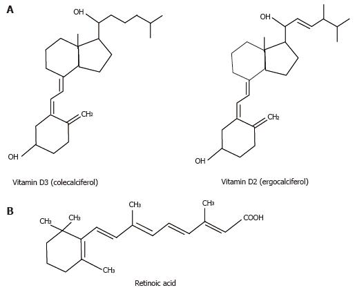
Figure 1 Structure of vitamin D and retinoic acid.
A: The chemical structure of vitamin D3 and vitamin D2 are indicated by line diagrams; B: The chemical structure of retinoic acid is indicated by line diagrams.
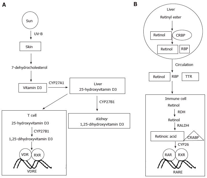
Figure 2 Vitamin D and retinoic acid biosynthesis and activity.
A: Vitamin D biosynthetic pathways are indicated, showing critical enzymes in particular tissues. Binding of VDR to DNA with RXR is indicated in a T cell; B: Retinoic acid biosynthetic pathways are indicated. Binding of RAR to DNA with RXR is indicated in an immune cell. RXR: Retinoid X receptor; VDR: Vitamin D receptor; RAR: Retinoic acid receptor; CRBP: Cellular retinol-binding proteins; RDH: Retinol dehydrogenases; VDRE: Vitamin D response element; RARE: Retinoic acid response element; CRABP: Cellular retinoic acid binding proteins.
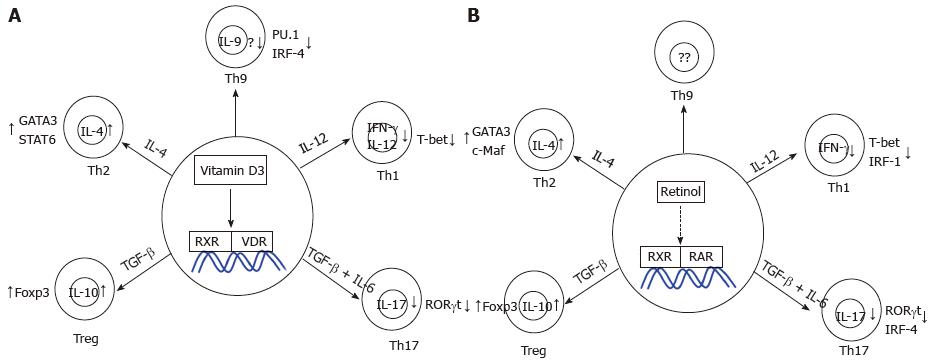
Figure 3 Effects of vitamin D and retinoic acid on T helper cell differentiation.
A: The effects of vitamin D on each of the T helper subsets are indicated, detailing changes in expression of cytokines and transcription factors; B: The effects of retinoic acid on each of the T helper subsets is indicated, detailing changes in expression of cytokines and transcription factors. IL: Interleukin; TGF: Transforming growth factor; Treg: Regulatory T cell.
- Citation: Goswami R, Kaplan MH. Essential vitamins for an effective T cell response. World J Immunol 2016; 6(1): 39-59
- URL: https://www.wjgnet.com/2219-2824/full/v6/i1/39.htm
- DOI: https://dx.doi.org/10.5411/wji.v6.i1.39


