Copyright
©The Author(s) 2017.
World J Clin Pediatr. Feb 8, 2017; 6(1): 103-109
Published online Feb 8, 2017. doi: 10.5409/wjcp.v6.i1.103
Published online Feb 8, 2017. doi: 10.5409/wjcp.v6.i1.103
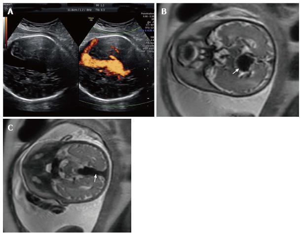
Figure 1 A 42-year-old G2P1001 presented for a routine growth ultrasound at 36 wk 5 d.
A: Prenatal ultrasonography shows vein of Galen aneurysm and color flow examination reveals a turbulence flow in the lesion and in the connected strait sinus; B: Prenatal T1-weighted magnetic resonance images show the markedly enlarged median procencephalic vein of Markowsky, characteristic of vein of Galen aneurysmal malformation (arrow); C: Persistence of the falcine draining sinus (arrow).
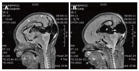
Figure 2 Sagittal T1-weighted magnetic resonance images show a choroidal type.
Vein of Galen aneurysmal malformation with feeders (arrow heads, A) to arteriovenous fistulas, dilated prosencephalic vein of Markowski and persistence of the falcine draining sinus (white arrow, B).
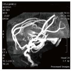
Figure 3 Magnetic resonance angiographic images show multiple enlarged arterial branches from the anterior and posterior cerebral arteries coalescing on the lateral margins of the dilated recipient vein.
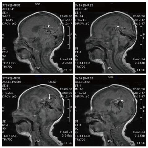
Figure 4 T1-weighted sagital magnetic resonance images at 41 d of age after 5 embolizations show thrombosis and shrinkage of the median vein of procencephalon and embryonic falcine sinus (arrows).
- Citation: Puvabanditsin S, Mehta R, Palomares K, Gengel N, Silva CFD, Roychowdhury S, Gupta G, Kashyap A, Sorrentino D. Vein of Galen malformation in a neonate: A case report and review of endovascular management. World J Clin Pediatr 2017; 6(1): 103-109
- URL: https://www.wjgnet.com/2219-2808/full/v6/i1/103.htm
- DOI: https://dx.doi.org/10.5409/wjcp.v6.i1.103


