Published online Feb 20, 2016. doi: 10.5321/wjs.v5.i1.15
Peer-review started: June 25, 2015
First decision: August 26, 2015
Revised: October 23, 2015
Accepted: December 1, 2015
Article in press: December 2, 2015
Published online: February 20, 2016
Processing time: 218 Days and 22.8 Hours
AIM: To analyze differences in prevalence and pattern of tooth agenesis and craniofacial morphology between non syndromic children with tooth agenesis with and without upper cervical spine morphological deviations and to analyze associations between craniofacial morphology and tooth agenesis in the two groups together.
METHODS: One hundred and twenty-six pre-orthodontic children with tooth agenesis were divided into two groups with (19 children, mean age 11.9) and without (107 children, mean age 11.4) upper spine morphological deviations. Visual assessment of upper spine morphology and measurements of craniofacial morphology were performed on lateral cephalograms. Tooth agenesis was evaluated from orthopantomograms.
RESULTS: No significant differences in tooth agenesis and craniofacial morphology were found between children with and without upper spine morphological deviations (2.2 ± 1.6 vs 1.94 ± 1.2, P > 0.05) but a tendency to a different tooth agenesis pattern were seen in children with morphological deviations in the upper spine. In the total group tooth agenesis was associated with the cranial base angle (n-s-ba, r = 0.23, P < 0.01), jaw angle (ML/RLar, r = 0.19, P < 0.05), mandibular inclination (NSL/ML, r = -0.21, P < 0.05), mandibular prognathia (s-n-pg, r = 0.25, P < 0.01), sagittal jaw relationship (ss-n-pg, r = -0.23, P < 0.5), overjet (r = -0.23, P < 0.05) and overbite (r = -0.25, P < 0.01).
CONCLUSION: Etiology of tooth agenesis in children with upper spine morphological deviations was discussed. The results may be valuable for the early diagnosis and treatment planning of non syndromic children with tooth agenesis.
Core tip: Tooth agenesis and craniofacial morphology was examined in non syndromic children with upper cervical spine morphological deviations. No significant differences in tooth agenesis and craniofacial morphology were found between children with and without upper spine morphological deviations, but a non-significant tendency of a different tooth agenesis pattern between the groups was seen. In the total group significant associations between tooth agenesis and craniofacial morphology were found. A different aetiology for tooth agenesis in children with morphological deviations in the upper spine was suggested. The results may be valuable for the early diagnosis and treatment planning of non syndromic children with tooth agenesis.
- Citation: Jasemi A, Sonnesen L. Tooth agenesis and craniofacial morphology in pre-orthodontic children with and without morphological deviations in the upper cervical spine. World J Stomatol 2016; 5(1): 15-21
- URL: https://www.wjgnet.com/2218-6263/full/v5/i1/15.htm
- DOI: https://dx.doi.org/10.5321/wjs.v5.i1.15
Tooth agenesis is a common congenital malformation that can occur either as an isolated finding or as part of a syndrome[1]. The complex and multifactoral etiology behind tooth agenesis is yet to be fully understood[2,3]. Tooth agenesis can occur as a result of mutations in genes involved in normal tooth development. Defects in the MSX-I and Sonic Hedgehog genes have been identified as causing tooth agenesis[2]. Furthermore, normal tooth development is dependent on the maturation of the bone surrounding the tooth germ and the nerve innervation of the teeth[3,4].
The prevalence of tooth agenesis among a healthy Danish population is between 7.8% and 8.2%[3,5]. Agenesis of the mandibular second premolar is most often observed (4.1%), followed by the maxillary second premolar (2.2%), the maxillary lateral incisors (1.7%) and the mandibular central incisors (0.2%)[6].
Previous studies have found an association between tooth agenesis and craniofacial morphology in non syndromic individuals[5,7-12]. It is generally agreed that tooth agenesis affects the craniofacial morphology in the sagittal and vertical dimension and that the deviation in the craniofacial morphology is associated with the prevalence and pattern of tooth agenesis[5,7-11]. In patients missing more than 12 teeth the prognathia of the mandible was more pronounced and the face was more squared compared to patients with less tooth agenesis[10].
The craniofacial morphology is also associated with upper cervical spine morphology in non syndromic individuals. In patients with severe skeletal malocclusion traits such as skeletal deep bite, skeletal open bite, skeletal maxillary and mandibular overjet, the prevalence of morphological deviations in the upper cervical spine was significantly higher compared to subjects with neutral occlusion and normal craniofacial morphology[13-16]. The pattern of morphological deviations in the upper cervical spine in these patients with severe skeletal malocclusions indicated fusion between the second and third cervical vertebra, block fusion between the second, third and fourth cervical vertebrae, occipitalization as assimilation of the first cervical vertebra with the occipital bone and partial cleft of the first cervical vertebra[13-16]. Furthermore, deviations of the upper cervical spine morphology were significantly associated with a large cranial base angle, retrognathia of the jaws and a large inclination of the jaws[13-16].
As associations between tooth agenesis and craniofacial morphology and associations between craniofacial morphology and upper cervical spine morphology have been described there may be an association between upper cervical spine morphology and tooth agenesis. To our knowledge the relation between upper cervical spine morphology and tooth agenesis has not yet been investigated.
Therefore, the aims of the present study are: (1) to analyze the differences in prevalence and pattern of tooth agenesis and craniofacial morphology between non syndromic children with tooth agenesis with and without upper cervical spine morphological deviations; and (2) to analyze the associations between craniofacial morphology and tooth agenesis in the two groups together.
The materials included cephalograms and orthopantomograms from non syndromic pre-orthodontic children registered between 1966 and 1997 at the orthodontic clinic, Muncipal Dental Service of Farum, Denmark. All the children with tooth agenesis that met the below inclusion criteria were included in the study: Children between 8 and 18 years old referred for orthodontic treatment before the orthodontic treatment began; one orthopantomogram and one lateral cephalogram before orthodontic treatment; agenesis of at least one permanent tooth, excluding the third molars; the first five cervical vertebrae visible on the lateral cephalogram. The exclusion criteria were: Children with known craniofacial or other syndromes; children with no tooth agenesis, excluding the thirds molars; children with insufficient medical records and X-rays.
A total of 126 children met these criteria and were included in the present study: 62 girls (aged 8-16 years, mean 11.32 years) and 64 boys (aged 8-16 years, mean 11.7 years) with an overjet ranging between -2.5 and 11 mm (mean 4.5 mm) and with an overbite ranging between -5 and 8 mm (mean 3.3 mm). According to the upper cervical spine morphology the children were divided into two groups: One group with upper cervical spine morphological deviations consisted of 19 children, 12 boys and 7 girls aged 9-14 years (mean age 11.9) and one group without upper cervical spine morphological deviations consisted of 107 children, 52 boys and 55 girls 8-16 years (mean age 11.4).
The study was approved by the Danish Data Protection Agency (No. 2013-54-0509).
Tooth agenesis was registered on orthopantomograms and the craniofacial and upper cervical spine morphology was registered on lateral cephalograms.
The registration of tooth agenesis was performed by visual assessment of the orthopantomograms. Only the permanent dentition was analyzed and the third molars were excluded from the study. Each registration on the orthopantomogram was compared with the individual child’s medical record and available information of the dentition. Only tooth agenesis where a tooth and its tooth bud was missing from the orthopantomogram and no history of extraction could be found in the corresponding medical record was registered. The registration included: Number of missing teeth; registration of multiple tooth agenesis (more than 4 missing teeth[10]); location of the tooth agenesis with regards to which jaw; agenesis pattern with regards to which tooth group (Tables 1 and 2).
| With upper spine deviations(n = 19) | Without upper spine deviations(n = 107) | Group | |||
| Variable | X-bar | SD | X-bar | SD | P |
| Age | 11.9 | 1.2 | 11.5 | 2 | NS |
| No. ageneses | 2.2 | 1.6 | 1.94 | 1.2 | NS |
| n-s-ar | 123.4 | 4 | 123.5 | 4.7 | NS |
| n-s-ba | 130 | 4.3 | 130.2 | 4.4 | NS |
| ML/RLar | 121 | 4.9 | 123.1 | 6.5 | NS |
| s-n-ss | 81.5 | 3.4 | 80.5 | 4 | NS |
| s-n-pg | 79.5 | 3.2 | 78.7 | 3.9 | NS |
| ss-n-pg | 2 | 1.7 | 1.8 | 2.6 | NS |
| NSL/NL | 8 | 2.7 | 7.1 | 3.6 | NS |
| NSL/ML | 30.4 | 4.5 | 31.6 | 5.6 | NS |
| NL/ML | 22.4 | 4.3 | 24.5 | 5.4 | NS |
| Overjet | 5 | 2.4 | 4.4 | 2.2 | NS |
| Overbite | 3.8 | 2 | 3.2 | 1.7 | NS |
| With upper spine deviations(n = 19) | Without upper spine deviations(n = 107) | Group | |||
| Variable | n | % | n | % | P |
| Gender | |||||
| Male | 12 | 63.2 | 52 | 48.6 | NS |
| Female | 7 | 36.8 | 55 | 51.4 | NS |
| Multiple ageneses | 1 | 5.3 | 5 | 4.7 | NS |
| Agenesis localization | |||||
| Mandible | 16 | 84.2 | 81 | 75.7 | NS |
| Maxilla | 8 | 42.1 | 52 | 48.6 | NS |
| Both jaws | 5 | 26.3 | 26 | 24.3 | NS |
| Agenesis tooth | |||||
| Incisor | 3 | 15.8 | 25 | 23.4 | NS |
| Canine | 0 | 0.00 | 2 | 1.9 | NS |
| Premolar | 17 | 89.5 | 85 | 79.4 | NS |
| Molar | 2 | 10.5 | 3 | 2.8 | NS |
| Several tooth groups | 3 | 15.8 | 7 | 6.5 | NS |
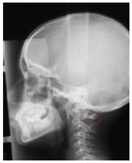
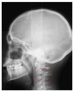
The cephalograms were studied for deviations in the morphology of the first five cervical vertebrae by visual assessment according to Sandham[17] and divided into two groups: Posterior arch deficiencies (PAD) and fusion anomalies. PAD consists of partial cleft and dehiscence. Partial cleft is defined as lack of fusion of the posterior arch[18] (Figure 1). Dehiscence is defined as inadequate development of a portion of the vertebra[18]. Fusion anomalies consist of fusion, block fusion and occipitalisation. Fusion is defined as fusion of two vertebrae at the articular facets, the posterior arch or the transverse process (Figure 2). Block Fusion is defined as fusion of more than two vertebrae at the vertebral bodies, the articular facets, the posterior arch or the transverse processes. Occipitalisation is defined as partial or complete fusion of the atlas (C1) with the occipital bone[17,18]. Morphological deviations were only registered if they were visible on all the cephalograms available in the medical record of the child. If, in the visual assessment of a cephalogram, any doubts occurred about the presence of morphological deviations, the subject was registered as having no morphological deviations in the upper spine. All cephalograms were reviewed together with supervisor LS.
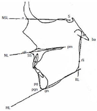
The craniofacial morphology was registered on lateral cephalograms of the children standing in the standardized head posture with their teeth in occlusion according to Siersbæk-Nielsen et al[19]. Twelve reference points were digitalized on cephalograms using the TIOPSTM software (Tiops 2005, Version 2.12.4) and nine angular measurements were measured according to Siersbæk-Nielsen et al[19]. Because the cephalograms were not scanned in a 1:1 scale, the overbite and overjet was measured by hand on analog cephalograms and taking into account the magnification of 5.6%. The points and lines are illustrated in Figure 3 and the mean values are shown in Table 1.
The reliability of the variables describing the cranial base and the vertical and sagittal craniofacial dimensions was assessed by re-measuring 25 lateral cephalograms selected at random from the previously evaluated cephalograms. The lateral cephalograms were marked and measured again, and paired t test found significant differences between the two sets of recordings related to the measurement of NSL/NL, NL/ML and ML/RLar. Since the pterygomaxillary point (Pm) is included in both NSL/NL and NL/ML, the location of the point was discussed and redefined. Subsequently, paired t test found no significant differences between the two sets of recordings. The method errors calculated by Dahlberg’s formula ranged from 0.01 to 1.32 degrees[20] and the Houston reliability coefficient from 0.89 to 1.00[21]. The reliability was within the average range as traditional film-based radiographs[22]. The reliability of the visual assessment of the morphological characteristics of the cervical vertebral units has previously been reported (k = 0.82)[23].
Regarding the craniofacial dimensions, the effect of age was assessed by linear regression analysis and for the occurrence of morphological deviations of the cervical column by logistic regression analysis. Differences in means of the craniofacial dimensions and number of tooth agenesis between genders and between the groups were assessed by unpaired t test. Differences in tooth agenesis pattern between genders and between the groups were assessed by Fisher’s exact test. Associations between tooth agenesis and craniofacial morphology and the possible effect of age and gender were tested by linear regression analyses. The results were considered significant at P values below 0.05. The statistical analyses were performed using SPSS 20.00 (Inc., Chicago, Illinois, United States).
No significant age and gender differences were found between children with and without morphological deviations in the upper cervical spine (Tables 1 and 2). In the group of children with morphological deviations in the upper spine (15.1% of the total group) the deviations occurred only as fusion between the second and third vertebra (42.3%) and partial cleft of the atlas (63.2%). Both morphological deviations occurred in 5.3% of the children with morphological deviations in the upper spine.
No statistically significant differences in tooth agenesis and craniofacial morphology were observed between children with and without morphological deviations in the upper spine. However, in children with morphological deviations in the upper spine a tendency to a different tooth agenesis pattern was seen as a larger occurrence of molar agenesis and agenesis of several tooth groups compared to the children without morphological deviations in the upper spine (Table 2).
In the total group, statistically significant associations were found between tooth agenesis and craniofacial morphology (Table 3). Multiple agenesis was positively associated with the gonial angle (ML/RLar; P <0.05) and significantly negatively associated with horizontal overjet (P < 0.05) and vertical overbite (P < 0.01; Table 3). Agenesis of incisors was negatively associated with the sagittal jaw relationship (ss-n-pg; P < 0.01). Agenesis of premolars was significantly positively associated with the cranial base angle (n-s-ba, P < 0.01) and the sagittal jaw relationship (ss-n-pg, P < 0.05; Table 3). Agenesis of the molars was significantly positively associated with the mandibular prognathia (s-n-pg, P < 0.01) and significantly negatively associated with the sagittal jaw relationship (ss-n-pg, P < 0.05) and the mandibular inclination (NSL/ML, P < 0.05; Table 3).
The present study has analyzed the differences in tooth agenesis and craniofacial morphology in pre-orthodontic children with tooth agenesis with and without upper cervical spine morphological deviations. To our knowledge this has not previously been reported in the literature. Additionally, the associations between tooth agenesis and craniofacial morphology in the two groups together were investigated.
In the total group of 126 non syndromic children with tooth agenesis, 15.1% had morphological deviations in the upper cervical spine which is in agreement with previous reported occurrence of morphological spine deviations in healthy adults with neutral occlusion, no tooth agenesis and normal craniofacial morphology (14.3%)[18]. Previous studies have shown that patients with severe skeletal malocclusions such as large overjet and overbite had a significantly higher occurrence of upper spine morphological deviations compared to controls[13-16]. Therefore a higher occurrence of morphological deviations in the upper spine was expected in the present study of children with tooth agenesis. One explanation for the relatively low occurrence of morphological deviations in the upper spine could be that the mean values for the overjet and overbite in the present study was within normal range and therefore children with severe malocclusion was few.
In the present study, no statistically significant differences in prevalence or pattern of tooth agenesis were found between non syndromic children with and without upper cervical spine morphological deviations. However, the non syndromic children with morphological deviations in the upper spine did show a tendency to have a greater percentage of molars agenesis and agenesis of several tooth groups compared to children without upper spine morphological deviations. In a healthy non syndromic population agenesis of the third molars are often seen, but agenesis of first and second molars as reported in children with morphological deviations in the upper spine in the present study almost never occurs[2,5] because normal tooth development is dependent on the maturation of the bone surrounding the tooth germ and the nerve innervation of the teeth[3,4]. Therefore it may be hypothesized that the etiology of tooth agenesis could be different in non syndromic children with morphological deviations in the upper spine as the tooth agenesis does not follow the normal pattern of tooth agenesis according to the nerve innervation. Previously, an association between the craniofacial skeleton and the upper cervical spine has been established[24-26]. An explanation for the association between the craniofacial skeleton including the jaws and teeth and the cervical spine could be found in the early embryogenesis. The notochord determines the development of the cervical spine, especially the vertebral bodies, and also the basilar part of the occipital bone in the cranial base which is the posterior part of the cranial base angle[27-33]. The para-axial mesoderm forming the vertebral arches and remaining parts of the occipital bone is also formed from the notochordal inductions. Therefore, a deviation in the development of the notochord may influence the surrounding bone tissue in the upper spine as well as the posterior part of the cranial base to which the jaws including the teeth are attached[24-26]. Only a non-significant tendency of differences in tooth pattern between children with and without morphological deviations in the upper cervical spine was found in the present study. This may be because the malocclusion and tooth agenesis were not extreme in the present sample and therefore a clear pattern could not be found.
Surprisingly, no statistically significant differences in the craniofacial morphology between the children with and without upper cervical spine morphological deviations were found. Previously, it has been shown that deviations of the upper cervical spine morphology were significantly associated with a large cranial base angle, retrognathia of the jaws and a large inclination of the jaws in non syndromic patients with severe skeletal malocclusion[13-16,18]. Therefore it was expected to find a difference in the craniofacial morphology between the two groups in the present study.
In agreement with previous studies[5,7-12] an association between tooth agenesis and the craniofacial morphology was found in the present study. In general, it was found that tooth agenesis was positively associated with the cranial base angle, gonial angle and the mandibular prognathia and negatively associated with the sagittal jaw relationship (except from agenesis of the premolars), mandibular inclination, overjet and overbite in the present study. The pattern of the association between the craniofacial morphology and tooth agenesis was in agreement with previous studies of non syndromic individuals[5,7-12].
In conclusion no significant differences in tooth agenesis and craniofacial morphology were found between the groups of children with and without morphological deviations in the upper spine, but a non-significant tendency to a different tooth agenesis pattern between the groups was seen. In the total group significant associations between tooth agenesis and craniofacial morphology were found. A different etiology for tooth agenesis in children with morphological deviations in the upper spine was suggested as these children may have a tendency for developing a different tooth agenesis pattern compared to children without upper spine morphological deviations. The results may be valuable in the early diagnosis and treatment planning of non syndromic children with tooth agenesis.
The Orthodontic clinic of the Municipal dental Service of Farum, Denmark is thanked for donating the material to the Department. Ib Jarle Christensen, Senior researcher, Department of Gastroenterology, Hvidovre Hospital, Denmark, is acknowledged for statistical advice. Copenhagen University Research Foundation (21-12-2012) is acknowledged for funding.
Associations between tooth agenesis and craniofacial morphology as well as associations between craniofacial morphology and upper cervical spine morphology have previously been found. The relation between upper cervical spine morphology and tooth agenesis has not yet been investigated.
Previously, an association between the craniofacial skeleton and the upper cervical spine has been established. An explanation for the association between the craniofacial skeleton including the jaws with the teeth and the cervical spine could be found in the early embryogenesis as a deviation in the development of the notochord.
As the relation between upper cervical spine morphology and tooth agenesis has not previously been investigated, the results of the present study may be a breakthrough in etiological and diagnostics considerations in non syndromic children with tooth agenesis.
Children with upper spine morphological deviations may have a tendency for developing a different tooth agenesis pattern compared to children without upper spine morphological deviations. Therefore a different etiology for tooth agenesis in children with morphological deviations in the upper spine was suggested. The results may be valuable in the early diagnosis and treatment planning of non syndromic children with tooth agenesis.
This is a report of a well conducted study. The pattern of tooth agenesis in patients with or without upper cervical spine anomalies was investigated and presented.
P- Reviewer: Cho SY, Gokul S, Rattan V, Vilchis R S- Editor: Ji FF L- Editor: A E- Editor: Wu HL
| 1. | Klein OD, Oberoi S, Huysseune A, Hovorakova M, Peterka M, Peterkova R. Developmental disorders of the dentition: an update. Am J Med Genet C Semin Med Genet. 2013;163C:318-332. [RCA] [PubMed] [DOI] [Full Text] [Cited by in Crossref: 75] [Cited by in RCA: 79] [Article Influence: 6.6] [Reference Citation Analysis (0)] |
| 2. | Kjær I, Becktor KB, Daugaard Jensen J, Russel B. Agenesi af tænder. Ny viden om forekomst og årsagsforhold. Tandlægebladet. 2000;104:198-202. |
| 3. | Kjaer I, Kocsis G, Nodal M, Christensen LR. Aetiological aspects of mandibular tooth agenesis--focusing on the role of nerve, oral mucosa, and supporting tissues. Eur J Orthod. 1994;16:371-375. [RCA] [PubMed] [DOI] [Full Text] [Cited by in Crossref: 47] [Cited by in RCA: 49] [Article Influence: 1.6] [Reference Citation Analysis (0)] |
| 4. | Miletich I, Sharpe PT. Normal and abnormal dental development. Hum Mol Genet. 2003;12 Spec No 1:R69-R73. [RCA] [PubMed] [DOI] [Full Text] [Cited by in Crossref: 119] [Cited by in RCA: 121] [Article Influence: 5.5] [Reference Citation Analysis (0)] |
| 5. | Rølling S. Hypodontia of permanent teeth in Danish schoolchildren. Scand J Dent Res. 1980;88:365-369. [PubMed] |
| 6. | Gungor AY, Turkkahraman H. Effects of severity and location of nonsyndromic hypodontia on craniofacial morphology. Angle Orthod. 2013;83:584-590. [RCA] [PubMed] [DOI] [Full Text] [Cited by in Crossref: 24] [Cited by in RCA: 26] [Article Influence: 2.2] [Reference Citation Analysis (0)] |
| 7. | Endo T, Yoshino S, Ozoe R, Kojima K, Shimooka S. Association of advanced hypodontia and craniofacial morphology in Japanese orthodontic patients. Odontology. 2004;92:48-53. [RCA] [PubMed] [DOI] [Full Text] [Cited by in Crossref: 43] [Cited by in RCA: 43] [Article Influence: 2.0] [Reference Citation Analysis (0)] |
| 8. | Tavajohi-Kermani H, Kapur R, Sciote JJ. Tooth agenesis and craniofacial morphology in an orthodontic population. Am J Orthod Dentofacial Orthop. 2002;122:39-47. [RCA] [PubMed] [DOI] [Full Text] [Cited by in Crossref: 71] [Cited by in RCA: 69] [Article Influence: 3.0] [Reference Citation Analysis (0)] |
| 9. | Wisth PJ, Thunhold K, Böe OE. The craniofacial morphology of individuals with hypodontia. Dentomaxillofac Radiol. 1975;4:70. [RCA] [PubMed] [DOI] [Full Text] [Cited by in Crossref: 2] [Cited by in RCA: 4] [Article Influence: 0.4] [Reference Citation Analysis (0)] |
| 10. | Nodal M, Kjaer I, Solow B. Craniofacial morphology in patients with multiple congenitally missing permanent teeth. Eur J Orthod. 1994;16:104-109. [RCA] [PubMed] [DOI] [Full Text] [Cited by in Crossref: 73] [Cited by in RCA: 75] [Article Influence: 2.4] [Reference Citation Analysis (0)] |
| 11. | Woodworth DA, Sinclair PM, Alexander RG. Bilateral congenital absence of maxillary lateral incisors: a craniofacial and dental cast analysis. Am J Orthod. 1985;87:280-293. [RCA] [PubMed] [DOI] [Full Text] [Cited by in Crossref: 58] [Cited by in RCA: 52] [Article Influence: 1.3] [Reference Citation Analysis (0)] |
| 12. | Zegan G, Mavru RB, Braha E. Craniofacial morphological changes of familial bilateral hypodontia of maxillary premolars. Rom J Morphol Embryol. 2014;55:443-448. [PubMed] |
| 13. | Sonnesen L, Kjaer I. Cervical column morphology in patients with skeletal Class III malocclusion and mandibular overjet. Am J Orthod Dentofacial Orthop. 2007;132:427.e7-427.12. [RCA] [PubMed] [DOI] [Full Text] [Cited by in Crossref: 44] [Cited by in RCA: 45] [Article Influence: 2.5] [Reference Citation Analysis (0)] |
| 14. | Sonnesen L, Kjaer I. Cervical vertebral body fusions in patients with skeletal deep bite. Eur J Orthod. 2007;29:464-470. [RCA] [PubMed] [DOI] [Full Text] [Cited by in Crossref: 48] [Cited by in RCA: 50] [Article Influence: 2.8] [Reference Citation Analysis (0)] |
| 15. | Sonnesen L, Kjaer I. Anomalies of the cervical vertebrae in patients with skeletal Class II malocclusion and horizontal maxillary overjet. Am J Orthod Dentofacial Orthop. 2008;133:188.e15-188.e20. [RCA] [PubMed] [DOI] [Full Text] [Cited by in Crossref: 35] [Cited by in RCA: 32] [Article Influence: 1.9] [Reference Citation Analysis (0)] |
| 16. | Sonnesen L, Kjaer I. Cervical column morphology in patients with skeletal open bite. Orthod Craniofac Res. 2008;11:17-23. [RCA] [PubMed] [DOI] [Full Text] [Cited by in Crossref: 40] [Cited by in RCA: 39] [Article Influence: 2.3] [Reference Citation Analysis (0)] |
| 17. | Sandham A. Cervical vertebral anomalies in cleft lip and palate. Cleft Palate J. 1986;23:206-214. [PubMed] |
| 18. | Sonnesen L. Associations between the Cervical Vertebral Column and Craniofacial Morphology. Int J Dent. 2010;2010:295728. [RCA] [PubMed] [DOI] [Full Text] [Full Text (PDF)] [Cited by in Crossref: 28] [Cited by in RCA: 26] [Article Influence: 1.7] [Reference Citation Analysis (0)] |
| 19. | Siersbaek-Nielsen S, Solow B. Intra- and interexaminer variability in head posture recorded by dental auxiliaries. Am J Orthod. 1982;82:50-57. [RCA] [PubMed] [DOI] [Full Text] [Cited by in Crossref: 90] [Cited by in RCA: 89] [Article Influence: 2.1] [Reference Citation Analysis (0)] |
| 20. | Dahlberg G. Statistical methods for medical and biological students. New York: Interscience Publications 1940; . |
| 21. | Houston WJ. The analysis of errors in orthodontic measurements. Am J Orthod. 1983;83:382-390. [RCA] [PubMed] [DOI] [Full Text] [Cited by in Crossref: 1085] [Cited by in RCA: 1098] [Article Influence: 26.1] [Reference Citation Analysis (0)] |
| 22. | Sonnesen L, Bakke M, Solow B. Temporomandibular disorders in relation to craniofacial dimensions, head posture and bite force in children selected for orthodontic treatment. Eur J Orthod. 2001;23:179-192. [RCA] [PubMed] [DOI] [Full Text] [Cited by in Crossref: 146] [Cited by in RCA: 140] [Article Influence: 5.8] [Reference Citation Analysis (0)] |
| 23. | Sonnesen L, Pedersen CE, Kjaer I. Cervical column morphology related to head posture, cranial base angle, and condylar malformation. Eur J Orthod. 2007;29:398-403. [RCA] [PubMed] [DOI] [Full Text] [Cited by in Crossref: 55] [Cited by in RCA: 52] [Article Influence: 2.9] [Reference Citation Analysis (0)] |
| 24. | Sonnesen L. Cervical vertebral column morphology associated with head posture and craniofacial morphology. Semin Orthod. 2012;18:118-125. [RCA] [DOI] [Full Text] [Cited by in Crossref: 7] [Cited by in RCA: 7] [Article Influence: 0.5] [Reference Citation Analysis (0)] |
| 25. | Sonnesen L, Nolting D, Kjaer KW, Kjaer I. Association between the development of the body axis and the craniofacial skeleton studied by immunohistochemical analyses using collagen II, Pax9, Pax1, and Noggin antibodies. Spine (Phila Pa 1976). 2008;33:1622-1626. [RCA] [PubMed] [DOI] [Full Text] [Cited by in Crossref: 16] [Cited by in RCA: 16] [Article Influence: 0.9] [Reference Citation Analysis (0)] |
| 26. | Sonnesen L, Nolting D, Engel U, Kjaer I. Cervical vertebrae, cranial base, and mandibular retrognathia in human triploid fetuses. Am J Med Genet A. 2009;149A:177-187. [RCA] [PubMed] [DOI] [Full Text] [Cited by in Crossref: 6] [Cited by in RCA: 6] [Article Influence: 0.4] [Reference Citation Analysis (0)] |
| 27. | Müller F, O’Rahilly R. The early development of the nervous system in staged insectivore and primate embryos. J Comp Neurol. 1980;193:741-751. [RCA] [PubMed] [DOI] [Full Text] [Cited by in Crossref: 24] [Cited by in RCA: 28] [Article Influence: 0.6] [Reference Citation Analysis (0)] |
| 28. | Kjaer I, Keeling JW, Graem N. Midline maxillofacial skeleton in human anencephalic fetuses. Cleft Palate Craniofac J. 1994;31:250-256. [RCA] [PubMed] [DOI] [Full Text] [Cited by in RCA: 1] [Reference Citation Analysis (0)] |
| 29. | Kjaer I. Human prenatal craniofacial development related to brain development under normal and pathologic conditions. Acta Odontol Scand. 1995;53:135-143. [RCA] [PubMed] [DOI] [Full Text] [Cited by in Crossref: 90] [Cited by in RCA: 92] [Article Influence: 3.1] [Reference Citation Analysis (0)] |
| 30. | Kjaer I. Neuro-osteology. Crit Rev Oral Biol Med. 1998;9:224-244. [RCA] [PubMed] [DOI] [Full Text] [Cited by in Crossref: 82] [Cited by in RCA: 83] [Article Influence: 3.1] [Reference Citation Analysis (0)] |
| 31. | Kjaer I, Fischer-Hansen B. The adenohypophysis and the cranial base in early human development. J Craniofac Genet Dev Biol. 1995;15:157-161. [PubMed] |
| 32. | Kjaer I, Niebuhr E. Studies of the cranial base in 23 patients with cri-du-chat syndrome suggest a cranial developmental field involved in the condition. Am J Med Genet. 1999;82:6-14. [RCA] [PubMed] [DOI] [Full Text] [Cited by in RCA: 2] [Reference Citation Analysis (0)] |
| 33. | Sadler TW. Embryology of neural tube development. Am J Med Genet C Semin Med Genet. 2005;135C:2-8. [RCA] [PubMed] [DOI] [Full Text] [Cited by in Crossref: 141] [Cited by in RCA: 134] [Article Influence: 6.7] [Reference Citation Analysis (0)] |


