Copyright
©The Author(s) 2022.
World J Obstet Gynecol. Nov 29, 2022; 11(4): 40-46
Published online Nov 29, 2022. doi: 10.5317/wjog.v11.i4.40
Published online Nov 29, 2022. doi: 10.5317/wjog.v11.i4.40
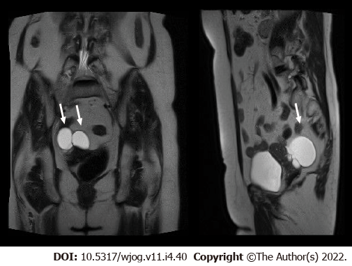
Figure 1 Magnetic resonance imaging scan showing the mass simulating an ovarian cyst (arrows).
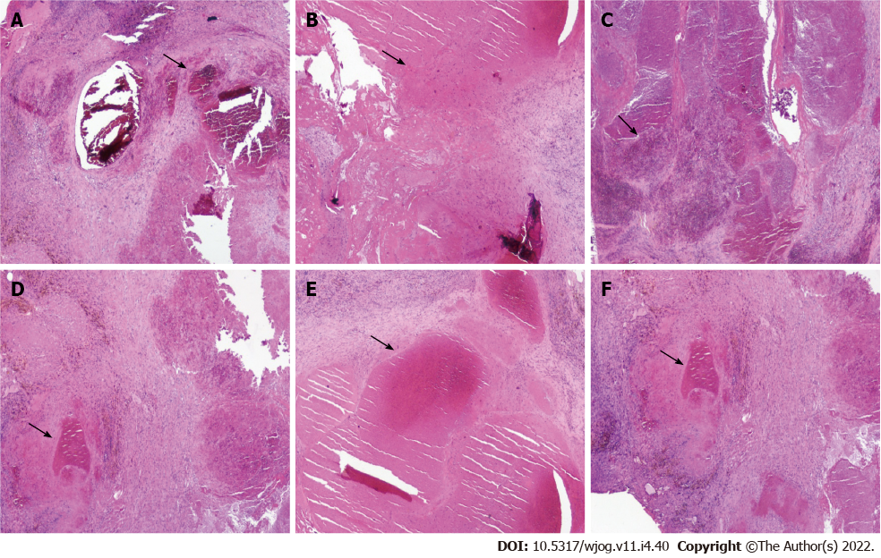
Figure 2 Histopathological analysis and immunohistochemical examination of the resected specimen.
A-F: Vascular lesion composed of circumscribed proliferation of blood vessels with different size and caliber. The thin of the wall is different in the different vassells. Necrosis and degenerative aspects are associated with lympho-monocitoid and plasmacell infiltration. The presence of macrophages with hemosiderin is evident (black arrow) (magnification 2×).
- Citation: Spinelli C, Ghionzoli M, Strambi S. Primary peritoneal hemangioendothelioma simulating an ovarian cyst: A case report and review of literature. World J Obstet Gynecol 2022; 11(4): 40-46
- URL: https://www.wjgnet.com/2218-6220/full/v11/i4/40.htm
- DOI: https://dx.doi.org/10.5317/wjog.v11.i4.40


