Copyright
©The Author(s) 2022.
World J Obstet Gynecol. May 20, 2022; 11(2): 8-16
Published online May 20, 2022. doi: 10.5317/wjog.v11.i2.8
Published online May 20, 2022. doi: 10.5317/wjog.v11.i2.8
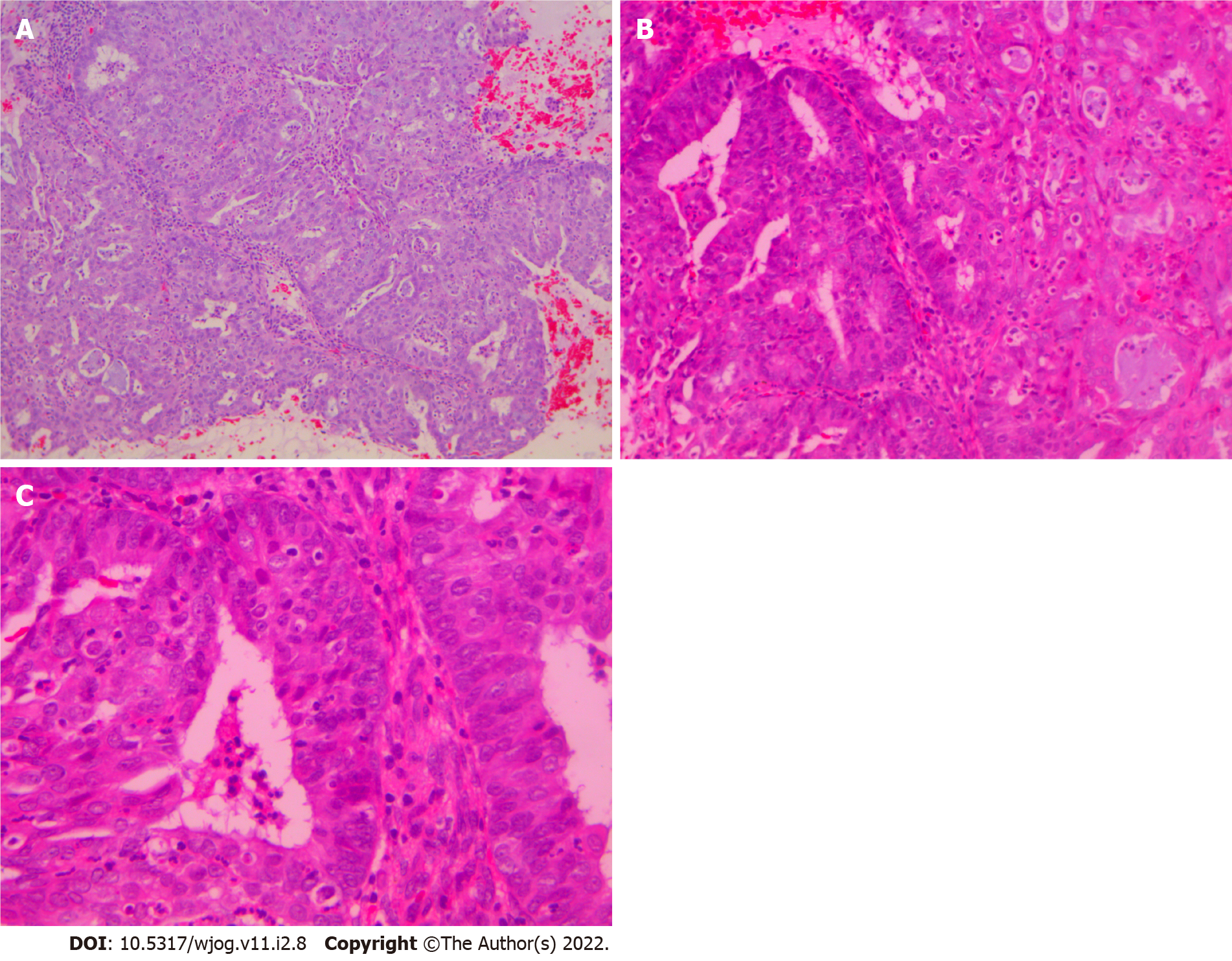
Figure 1 Microglandular hyperplasia-like pattern in dilatation and curettage, hematoxylin-eosin staining.
A: Complex microglandular proliferation of small back-to-back glands, without intervening stroma, with mucin production and accumulation of neutrophils in the gland lumen and stroma (× 10); B: Rare area where Microglandular hyperplasia-like adenocarcinoma (on the right) is merging with more conventional endometrioid adenocarcinoma (on the left) (× 20); C: Endometrioid adenocarcinoma area shows pseudostratification, mild atypia and rare mitoses (× 40).
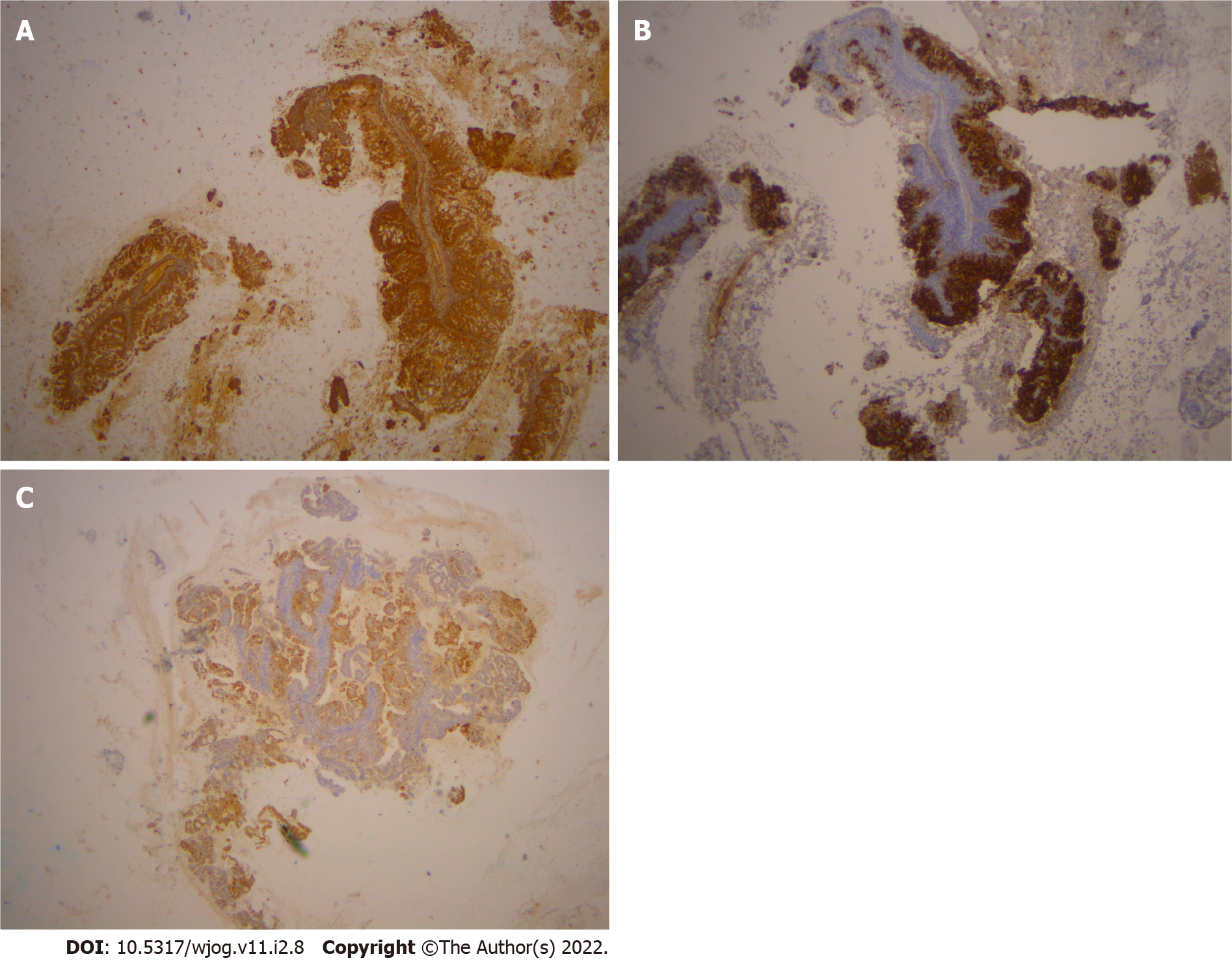
Figure 2 Immunohistochemical staining.
A: Immunohistochemical stain for VIM shows positive stain in the microglandular hyperplasia-like adenocarcinoma (MGA) in the curettage specimen (× 10); B: Immunohistochemical stain for CEA shows positive stain in the MGA in the curettage specimen (× 10); C: Immunohistochemical stain shows positive expression for p16 in the MGA in the curettage specimen (× 10).
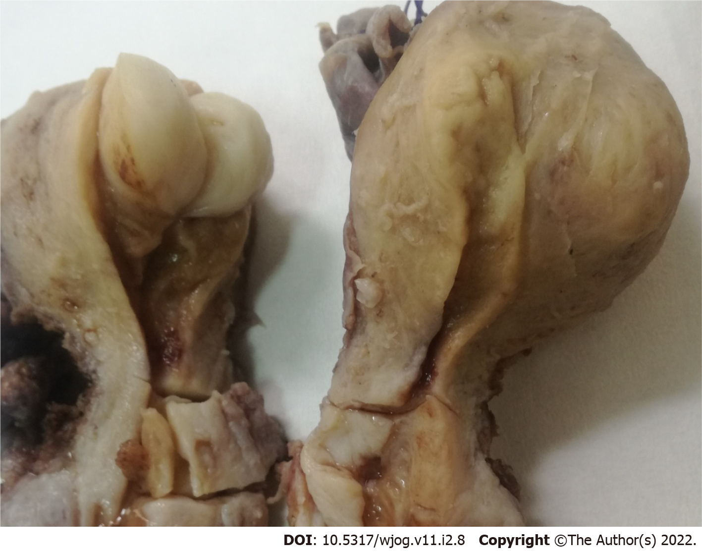
Figure 3 Macroscopic appearance of the uterus.
There are obvious the leiomyoma, the cervical polyp and the flat appearance of the endometrial area.
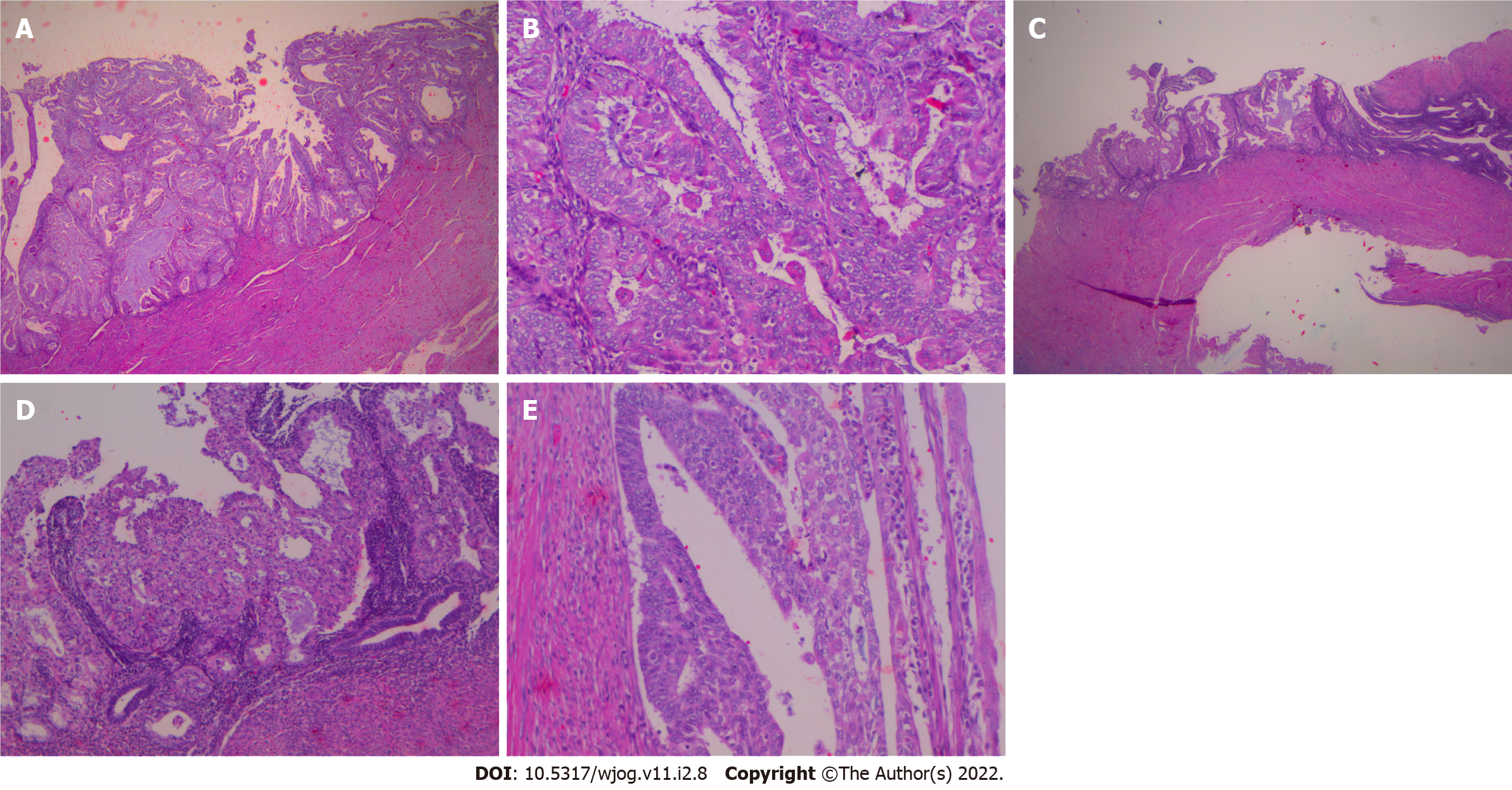
Figure 4 Hysterectomy specimen.
A: Residual carcinoma of conventional mucinous type in the hysterectomy specimen (× 20); B: Areas of ciliated and small non-villous papillae in the residual adenocarcinoma of the endometrium in the hysterectomy specimen (× 20); C: Adenocarcinoma with microglandular hyperplasia (MGH)-like pattern occurs on the tumor surface of conventional adenocarcinoma in a plaque-like fashion (× 10); D: Higher magnification of C (× 20); E: Adenocarcinoma with MGH-like pattern occurs on the tumor surface of conventional adenocarcinoma (another area) (× 40).
- Citation: Trihia HJ, Souka E, Galanopoulos G, Pavlakis K, Karelis L, Fotiou A, Provatas I. Microglandular hyperplasia-like mucinous adenocarcinoma of the endometrium: A rare case report. World J Obstet Gynecol 2022; 11(2): 8-16
- URL: https://www.wjgnet.com/2218-6220/full/v11/i2/8.htm
- DOI: https://dx.doi.org/10.5317/wjog.v11.i2.8


