Copyright
©The Author(s) 2022.
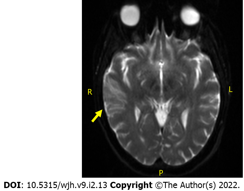
Figure 1 Diffusion-weighted magnetic resonance imaging of right middle cerebral artery watershed embolic infarct.
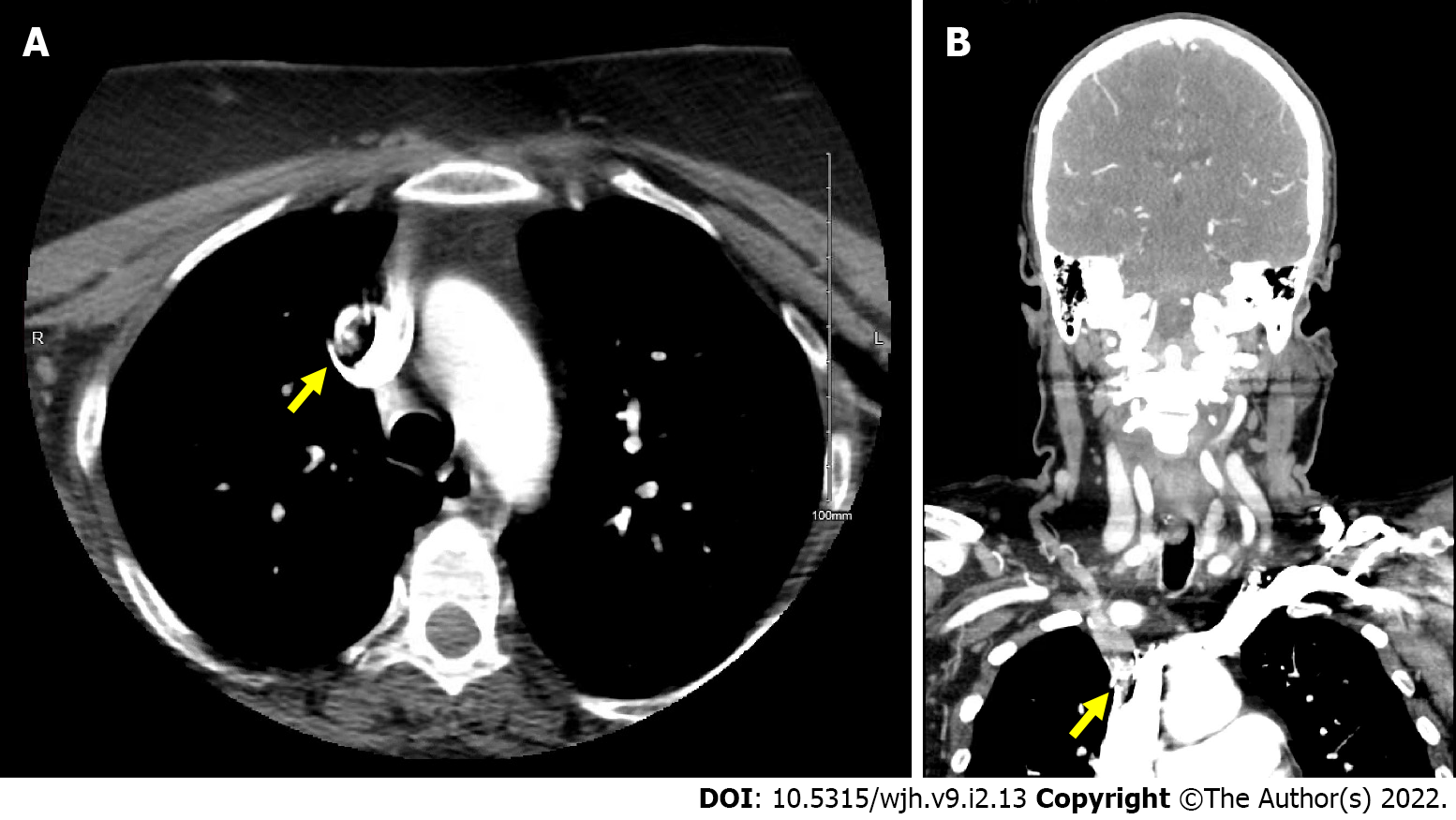
Figure 2 Computed tomography image.
A: Head and Neck computed tomography angiogram in transverse section with filling defect noted in base of proximal right brachiocephalic artery. B: Computed tomography angiogram in coronal section with filling defect noted in base of proximal right brachiocephalic artery.
- Citation: Kilby KJ, Anderson-Quiñones C, Pierce KR, Gabrah K, Seth A, Brunson A. Late ischemic stroke and brachiocephalic thrombus in a 65-year-old patient six months after COVID-19 infection: A case report. World J Hematol 2022; 9(2): 13-19
- URL: https://www.wjgnet.com/2218-6204/full/v9/i2/13.htm
- DOI: https://dx.doi.org/10.5315/wjh.v9.i2.13


