Copyright
©The Author(s) 2017.
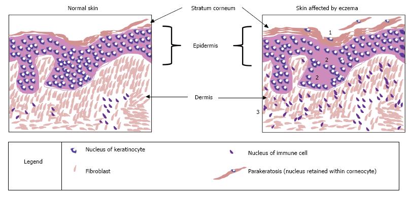
Figure 2 Histological features of atopic eczema.
This figure represents sections of skin biopsies, stained with haematoxylin and eosin, to highlight the changes that can be observed under a light microscope, comparing skin affected by atopic eczema and a control sample. Three characteristic features of atopic eczema are illustrated: Increased thickness of the stratum corneum (hyperkeratosis and parakeratosis) is caused by a disruption to the cornification process; spongiosis occurs when there is a reduction or damage of the proteins involved in tight junctions, thereby leading to uncontrolled movement of fluids in the paracellular space; infiltration of the dermis by immune cells is a sign of the immune response as a primary feature of atopic eczema itself or in response to the entry of allergens through a leaky skin barrier. The effects of eczema: 1: Increased thickness of stratum corneum; 2: Spongiosis - oedema (water retained between cells); 3: Increased number of immune cells.
- Citation: Bell DC, Brown SJ. Atopic eczema treatment now and in the future: Targeting the skin barrier and key immune mechanisms in human skin. World J Dermatol 2017; 6(3): 42-51
- URL: https://www.wjgnet.com/2218-6190/full/v6/i3/42.htm
- DOI: https://dx.doi.org/10.5314/wjd.v6.i3.42


