Copyright
©The Author(s) 2017.
Figure 1 Histopathological examination.
A: Foam cells infiltrate the superficial and deep dermis in cluster, separated by collagen fibers. Absence of any other significant inflammatory infiltrate (10 ×, EE); B: Xanthoma cells are filled with optically empty vacuoles, showing thin, well defined cytoplasmic membranes, and tend to be attached to each other. They can be multinucleate (20 ×, EE); C: Presence in the dermis of cholesterol crystalline aggregates surrounded by fibrosis and foamy cells (10 ×, EE).
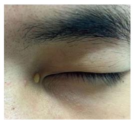
Figure 2 Xanthelasma.
Single yellow-orange papular lesion on the inner canthus of the eye.
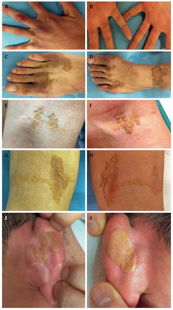
Figure 3 Diffuse intertriginous xanthomas.
Usually appear in a symmetric distribution as well-demarcated and slightly elevated noninflammatory plaques of ochre-yellow or yellow-brown discoloration. Typically found in intertriginous and flexural areas. A: In finger web spaces, and in this picture with metacarpophalangeal joint tendon xanthoma; B: At metacarpophalangeal palmar crease in linear band or single papules; C: Toe web spaces; D: In toe web spaces and ankle crease; E and F: At antecubital fossae, with the “eruptive” appearance of crops of yellow dermal soft, velvety papules; G and H: In popliteal fossae; I and J: At the creases of ears in a rare pattern of “plane xanthoma” as very thin flat patches, easily clinically missed, of yellow-orange macular discoloration.
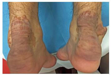
Figure 4 Tendinous xanthomas.
Bilateral Xanthomas of Achilles tendon. Each swelling was localized all over the tendon just above its insertion point to the calcaneal tuberosity. They appear as firm, mobile, painless slowly enlarging subcutaneous nodules which may join together to form a single mass or multilobated masses. They are covered by reddish-brown thickened skin.
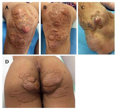
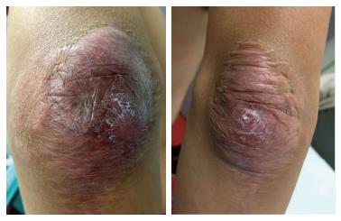
Figure 5 Tuberous xanthomas.
They are very common and clinically variable. They may appear as firm, painless, red-yellow, waxy-appearing nodules located in the dermis and subcutaneous tissue, from few millimeters to several centimeters in size. They often present with a cobblestone-like pattern developing around the pressure areas such as: A and B: The knees; C: Malleolus; D: Buttocks. Lesions can join together to form multilobated masses.
Figure 6 Bilateral massive tuberous xanthomas of the elbows did not recur after surgical excision.
- Citation: Mastrolorenzo A, D’Errico A, Pierotti P, Vannucchi M, Giannini S, Fossi F. Pleomorphic cutaneous xanthomas disclosing homozygous familial hypercholesterolemia. World J Dermatol 2017; 6(4): 59-65
- URL: https://www.wjgnet.com/2218-6190/full/v6/i4/59.htm
- DOI: https://dx.doi.org/10.5314/wjd.v6.i4.59


