Copyright
©The Author(s) 2017.
World J Orthop. Mar 18, 2017; 8(3): 256-263
Published online Mar 18, 2017. doi: 10.5312/wjo.v8.i3.256
Published online Mar 18, 2017. doi: 10.5312/wjo.v8.i3.256
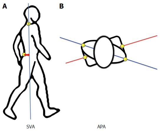
Figure 2 Sagittal vertical axis and angle pelvis-acromion.
A: SVA was defined as the distance between the marker “S1” and the vertical line passing by the marker “C7”. This parameter reflects trunk position during gait: A great value of SVA indicates that the trunk is leaning forward; B: APA was defined as the angle between the line joining the 2 “Acromion” markers and the line joining the 2 “anterosuperior iliac spine” markers. SVA: Sagittal vertical axis; APA: Angle pelvis-acromion.
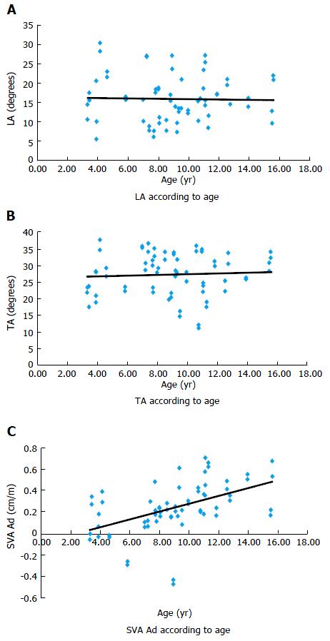
Figure 3 Continuous analysis of kinematic parameters according to the age.
A: TA; B: LA; C: SVA. TA: Thoracic angle; LA: Lumbar angle; SVA: Sagittal vertical axis.
Figure 4 Continuous analysis of angle pelvis-acromion-rom according to the age.
APA: Angle pelvis-acromion.
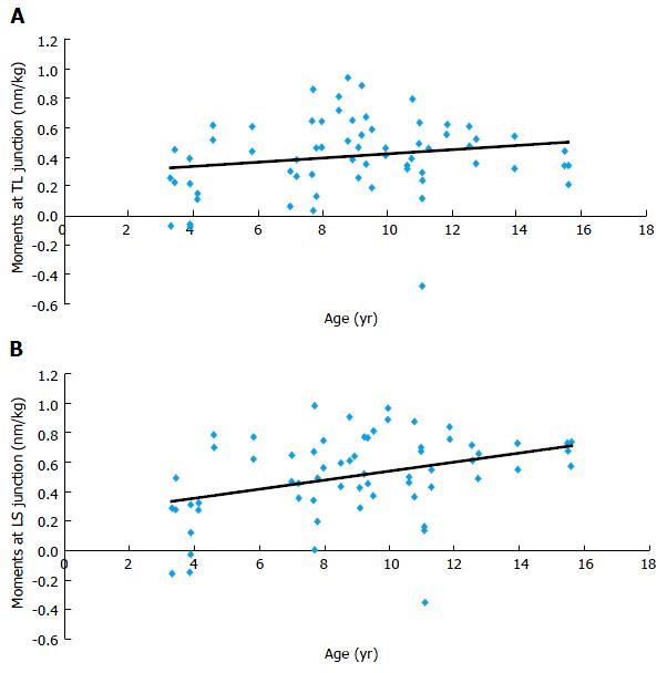
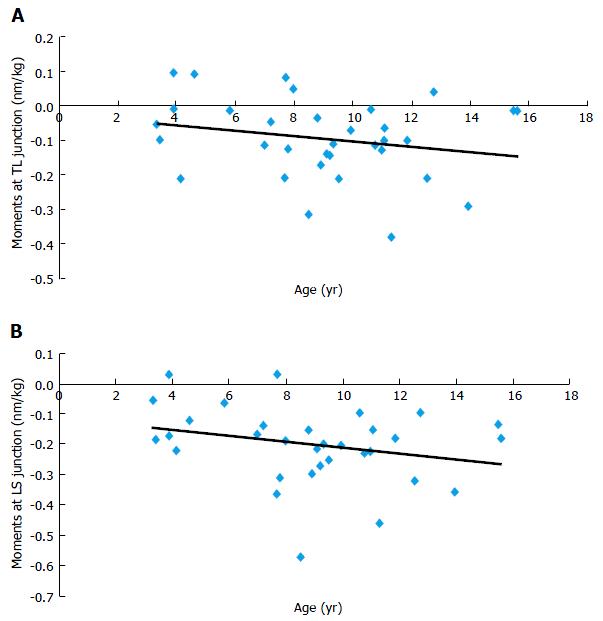
Figure 5 Sagittal kinetic parameters of the trunk according to the age.
A: TL; B: LS. Frontal plane constraints are relative to flexion-extension movements. TL: Thoracolumbar; LS: Lumbosacral.
Figure 6 Transversal kinetic parameters of the trunk according to the age (continuous analysis).
Transversal plane constraints are relative to torsional movements of the trunk. TL: Thoracolumbar; LS: Lumbosacral.
- Citation: Pesenti S, Blondel B, Peltier E, Viehweger E, Pomero V, Authier G, Fuentes S, Jouve JL. Spinal alignment evolution with age: A prospective gait analysis study. World J Orthop 2017; 8(3): 256-263
- URL: https://www.wjgnet.com/2218-5836/full/v8/i3/256.htm
- DOI: https://dx.doi.org/10.5312/wjo.v8.i3.256


