Copyright
©The Author(s) 2017.
World J Orthop. Oct 18, 2017; 8(10): 770-776
Published online Oct 18, 2017. doi: 10.5312/wjo.v8.i10.770
Published online Oct 18, 2017. doi: 10.5312/wjo.v8.i10.770
Figure 1 Preoperative radiograph showing retropharyngeal/prevertebral soft tissue at the level of C2 vertebral body and at the level of C6 vertebral body.
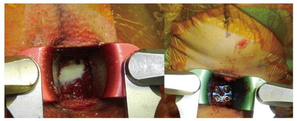
Figure 2 Photograph showing demineralized bone matrix packed within cage after insertion into disc space and anterior cervical discectomy and fusion plate.
Figure 3 Plain lateral radiograph of the cervical spine, taken one week postoperatively, which shows the normal dimensions of the prevertebral space being less than 50% of the vertebral body at C2 and less than the body width of C6 respectively.
- Citation: Chin KR, Pencle FJR, Seale JA, Valdivia JM. Soft tissue swelling incidence using demineralized bone matrix in the outpatient setting. World J Orthop 2017; 8(10): 770-776
- URL: https://www.wjgnet.com/2218-5836/full/v8/i10/770.htm
- DOI: https://dx.doi.org/10.5312/wjo.v8.i10.770


