Copyright
©2013 Baishideng Publishing Group Co.
World J Orthop. Oct 18, 2013; 4(4): 229-240
Published online Oct 18, 2013. doi: 10.5312/wjo.v4.i4.229
Published online Oct 18, 2013. doi: 10.5312/wjo.v4.i4.229
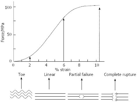
Figure 1 Stress-strain relationship for progressive loading of a tendon showing three distinct regions (toe, linear and partial failure) prior to complete rupture approximate stress forces (MPa) and strain values (% strain) is shown.
Reprinted from [40] with permission from the Oxford University Press.
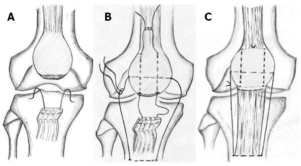
Figure 2 Drawing illustrates repair of the patellar tendon and making of the “suture line tension regulating suture”.
A: The modified Kessler suture in the proximal portion of an avulsed patellar tendon; B: The modified Kessler suture and the reinforcement device before tying the threads into a knot; C: The final appearance after tying the threads into knots. Reprinted from [124] with permission from the Springer.
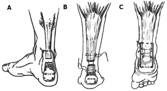
Figure 3 Drawing illustrates repair of the ruptured Achilles tendon.
A: Thread of the reinforcement suture when it passes through the osseous tunnel in the calcaneus and by interweaving through the proximal stump of the Achilles tendon; B: Tendon stumps that were held together by Kessler’s suture; C: Final appearance of the reinforcement suture and the repaired Achilles tendon. Reprinted from [125] with permission from the Elsevier.
- Citation: Massoud EIE. Healing of subcutaneous tendons: Influence of the mechanical environment at the suture line on the healing process. World J Orthop 2013; 4(4): 229-240
- URL: https://www.wjgnet.com/2218-5836/full/v4/i4/229.htm
- DOI: https://dx.doi.org/10.5312/wjo.v4.i4.229


