Copyright
©The Author(s) 2025.
World J Orthop. Jan 18, 2025; 16(1): 101656
Published online Jan 18, 2025. doi: 10.5312/wjo.v16.i1.101656
Published online Jan 18, 2025. doi: 10.5312/wjo.v16.i1.101656
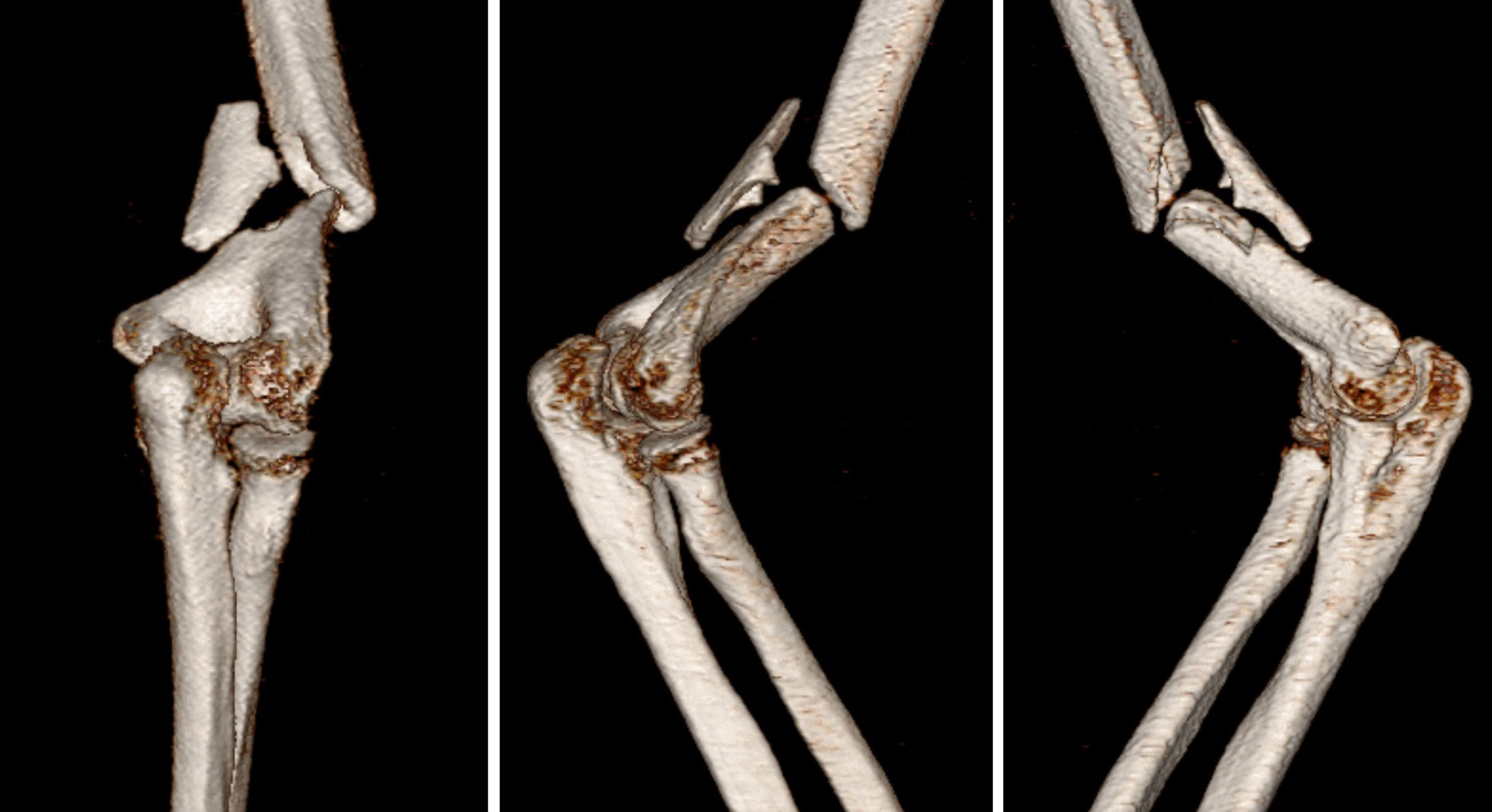
Figure 1 Preoperative three-dimensional computed tomography imaging conducted upon hospital admission.
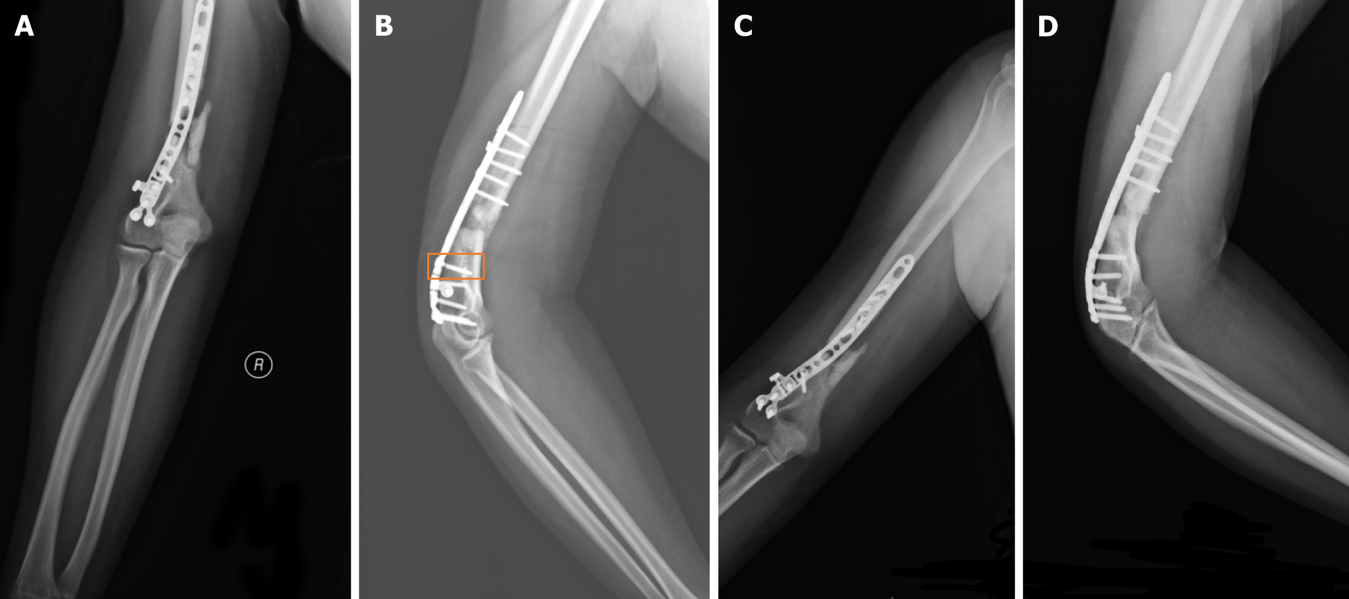
Figure 2 X-ray images.
A: X-ray images taken four months after surgery anterior-posterior view; B: X-ray images taken four months after surgery lateral view. The orange box highlights the location of the broken screw; C: X-ray images taken nine months after surgery anterior-posterior view; D: X-ray images taken nine months after surgery lateral view.
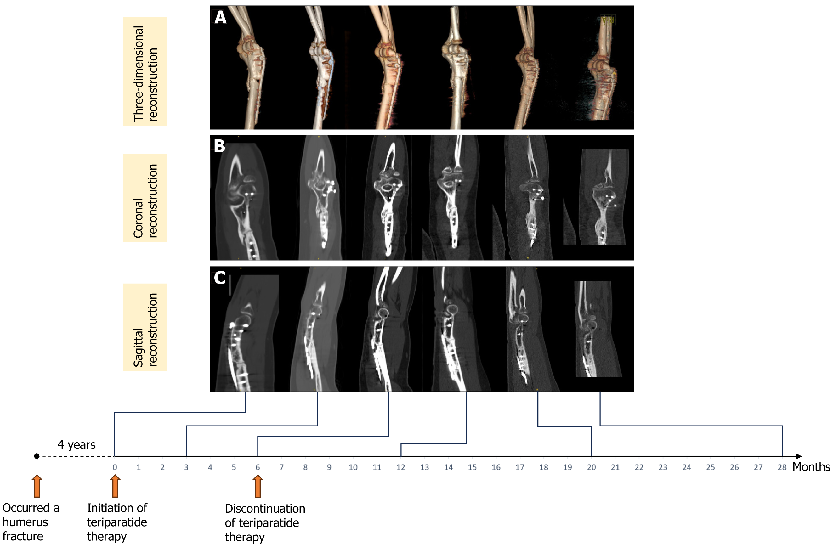
Figure 3 Computed tomography scan images showing the progression of fracture healing before and after teriparatide treatment.
A: Three-dimensional reconstruction; B: Coronal two-dimensional reconstruction; C: Sagittal two-dimensional reconstruction. From left to right: Prior to teriparatide treatment (four years after the initial fracture surgery), 3 months, 6 months, 12 months, 20 months and 28 months post-teriparatide treatment initiation. Note: The teriparatide treatment duration was 6 months.
- Citation: Guo SH, Li C, Gao YJ, Zhang Z, Lu K. Teriparatide as a non-surgical salvage therapy for prolonged humerus fracture nonunion: A case report and literature review. World J Orthop 2025; 16(1): 101656
- URL: https://www.wjgnet.com/2218-5836/full/v16/i1/101656.htm
- DOI: https://dx.doi.org/10.5312/wjo.v16.i1.101656


