Copyright
©The Author(s) 2024.
World J Orthop. Aug 18, 2024; 15(8): 813-819
Published online Aug 18, 2024. doi: 10.5312/wjo.v15.i8.813
Published online Aug 18, 2024. doi: 10.5312/wjo.v15.i8.813
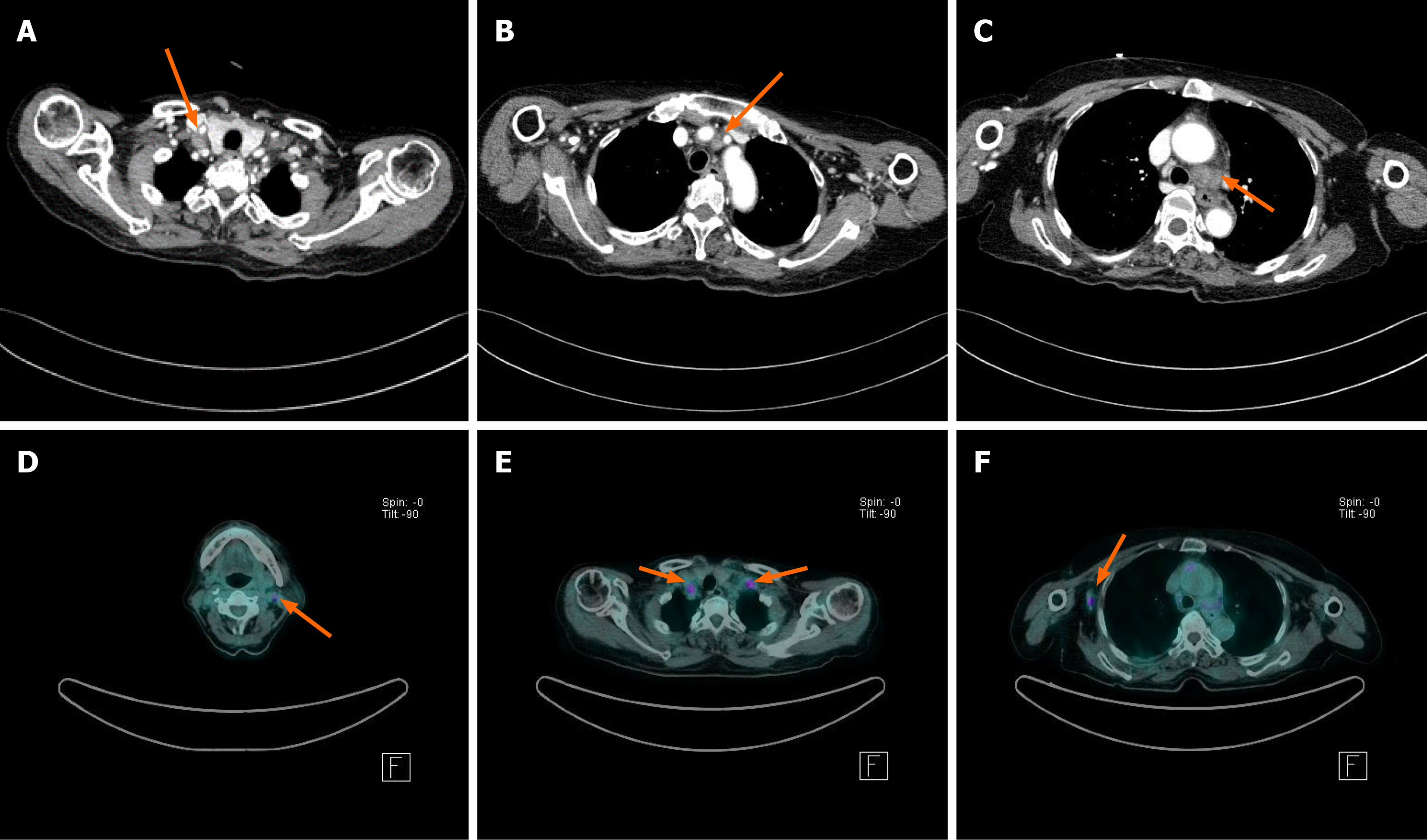
Figure 1 Computed tomography and fluorine-2-fluoro-2-deoxy-d-glucose positron emission tomography/computerized tomography findings.
A-C: Enlargement of both supraclavicular and mediastinal lymph nodes was detected via computed tomography (CT); D-F: Positron emission tomography/CT revealed 18F-fluorodeoxyglucose-uptake in the right cervical, both supraclavicular, and left axillary lymph nodes.
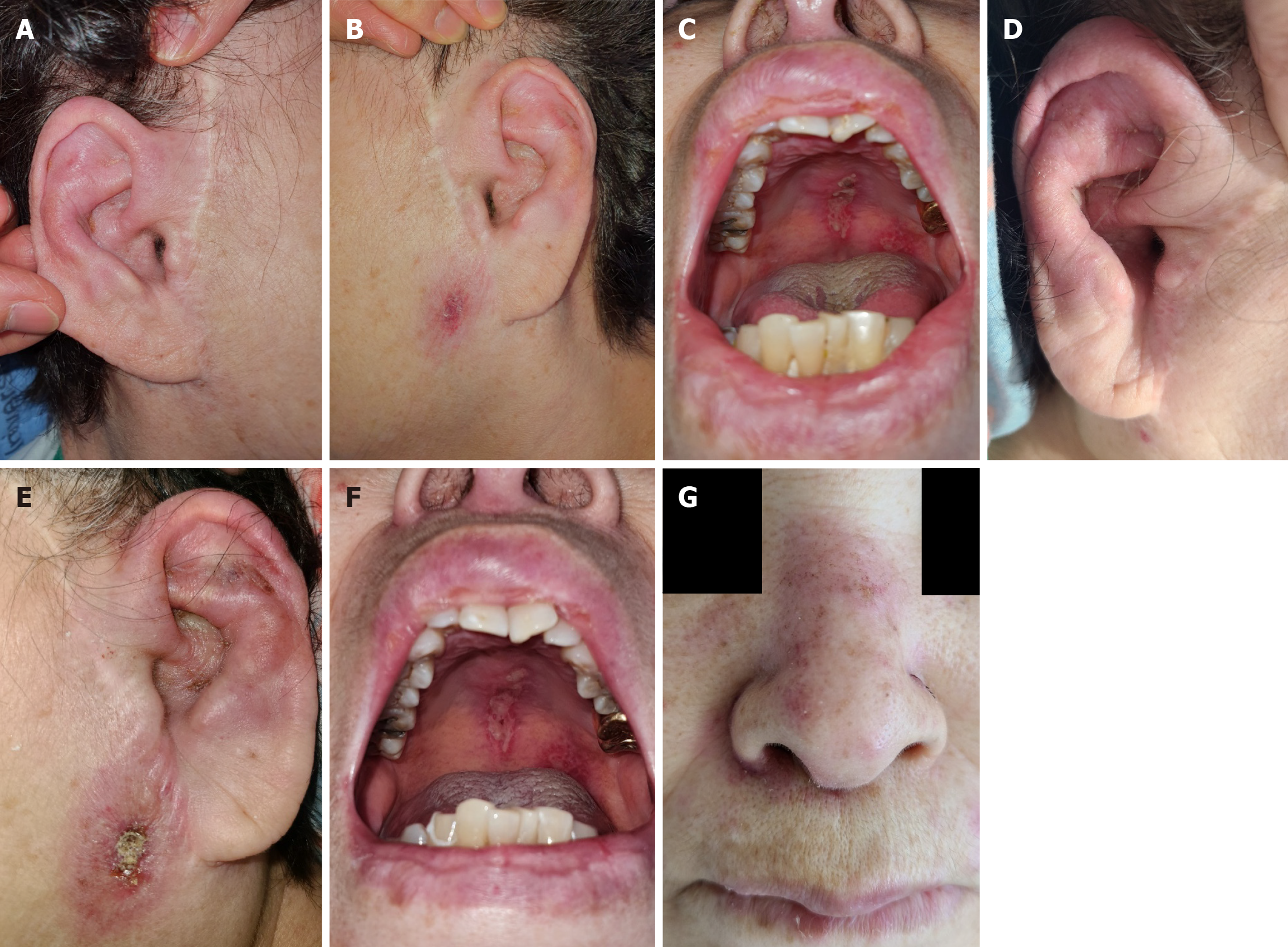
Figure 2 Photographs of a physical examination.
A-C: Erythema and mild swelling on both auricles and ulcerative mucosal lesions on palates on the second day of admission; D-G: On day 10, erythema and swelling intensified on both auricles, with increased periauricular skin inflammation on the left side, and oral mucosal ulceration worsened, and erythema appeared on the nasal ridge.
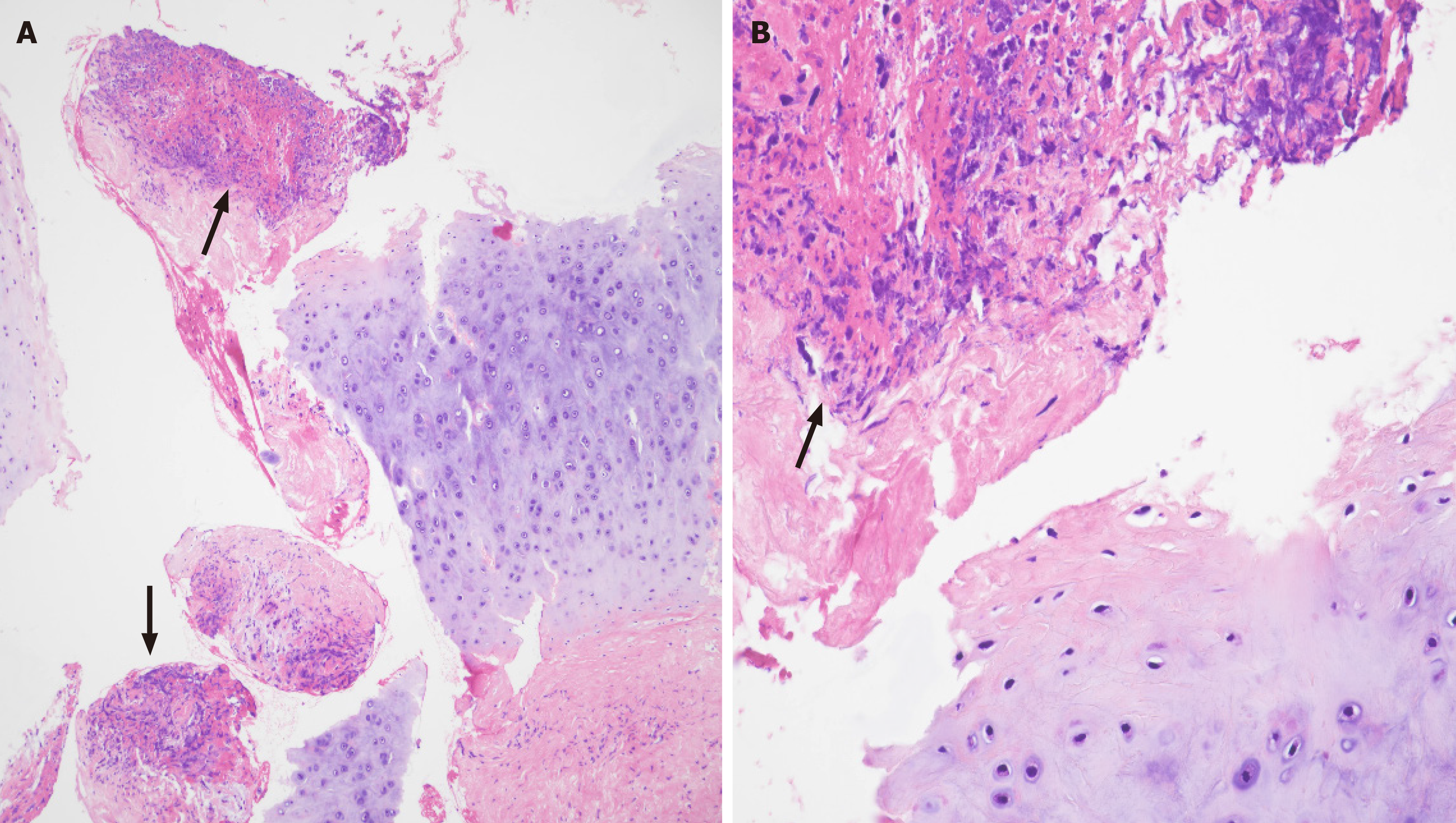
Figure 3 Histologic findings.
Histologic findings of relapsing polychondritis of the ear. The fragmented biopsy specimen containing ear cartilage and adjacent fibrous tissue, with inflammatory infiltration and necrosis (arrows). A: Original magnification × 100; B: Original magnification × 400.
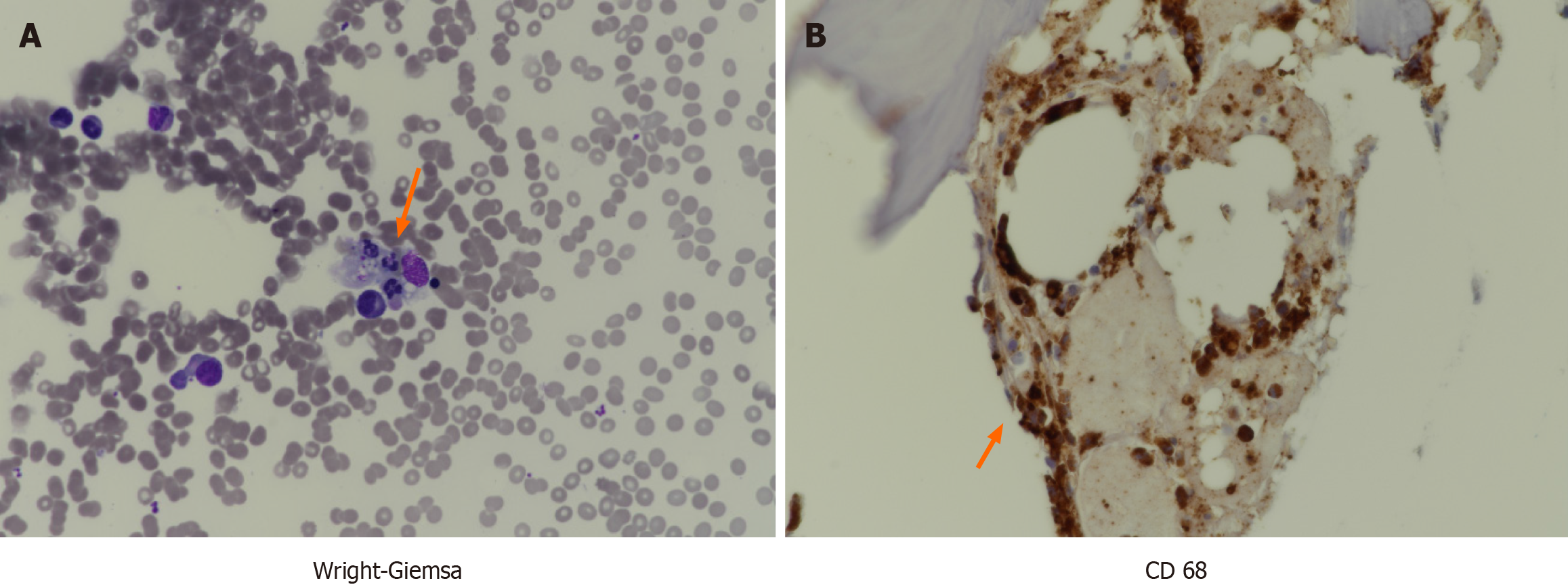
Figure 4 Bone marrow findings.
A: Phagocytosis of blood cells is observed in the bone marrow stained with Wright-Giemsa (arrow); B: Cluster of differentiation 68 [CD68 (+)], a protein primarily expressed in cells such as macrophages, suggests excessive cellular activation and an inflammatory response.
- Citation: Han MR, Hwang JH, Cha S, Jeon SY, Jang KY, Kim N, Lee CH. Hemophagocytic lymphohistiocytosis triggered by relapsing polychondritis: A case report. World J Orthop 2024; 15(8): 813-819
- URL: https://www.wjgnet.com/2218-5836/full/v15/i8/813.htm
- DOI: https://dx.doi.org/10.5312/wjo.v15.i8.813


