Copyright
©The Author(s) 2024.
World J Orthop. Nov 18, 2024; 15(11): 1095-1100
Published online Nov 18, 2024. doi: 10.5312/wjo.v15.i11.1095
Published online Nov 18, 2024. doi: 10.5312/wjo.v15.i11.1095
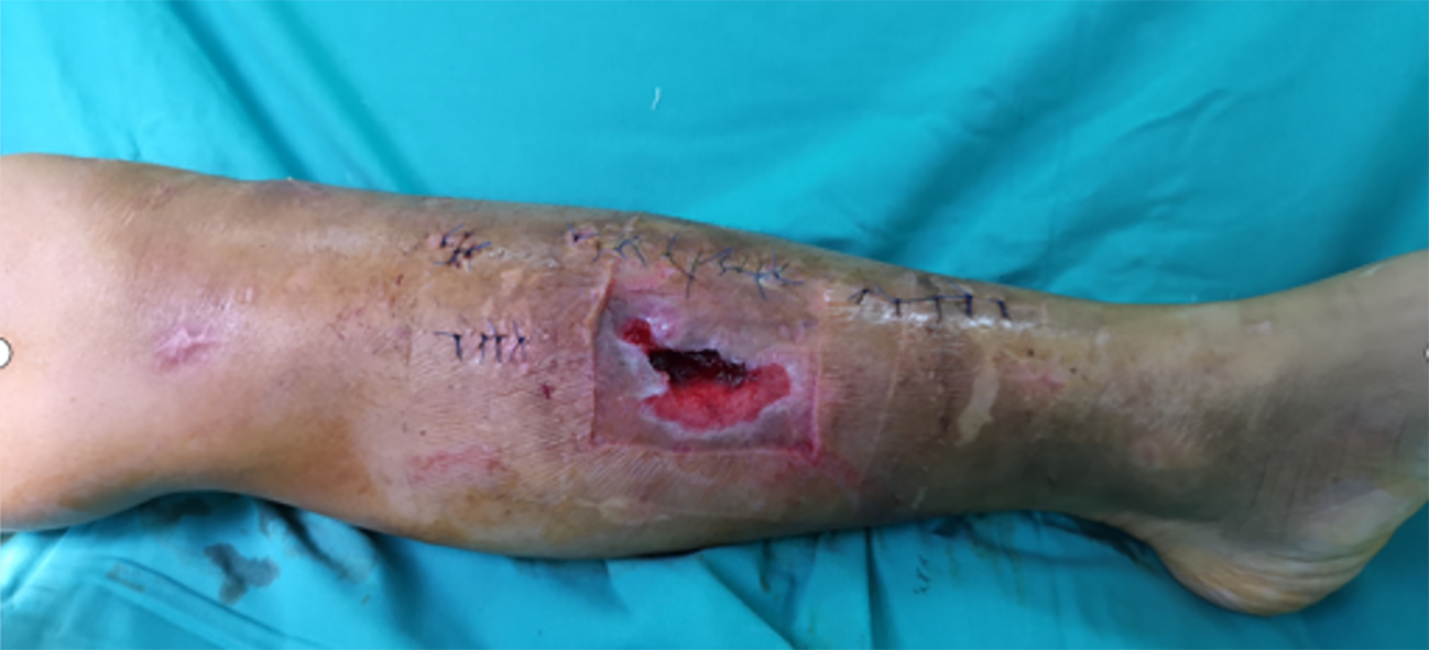
Figure 1 Soft tissue lesions.
The wound defect was dark red, with mild redness and swelling of the surrounding skin. Local skin defects have irregular shapes with uneven edges. The wound defect was about 3 centimeters long, 2 centimeters wide, and up to 0.5 centimeters deep. Moderate swelling was observed around the wound, with disappearance of local skin lines. The skin around the wound felt numb and the pain was dull.
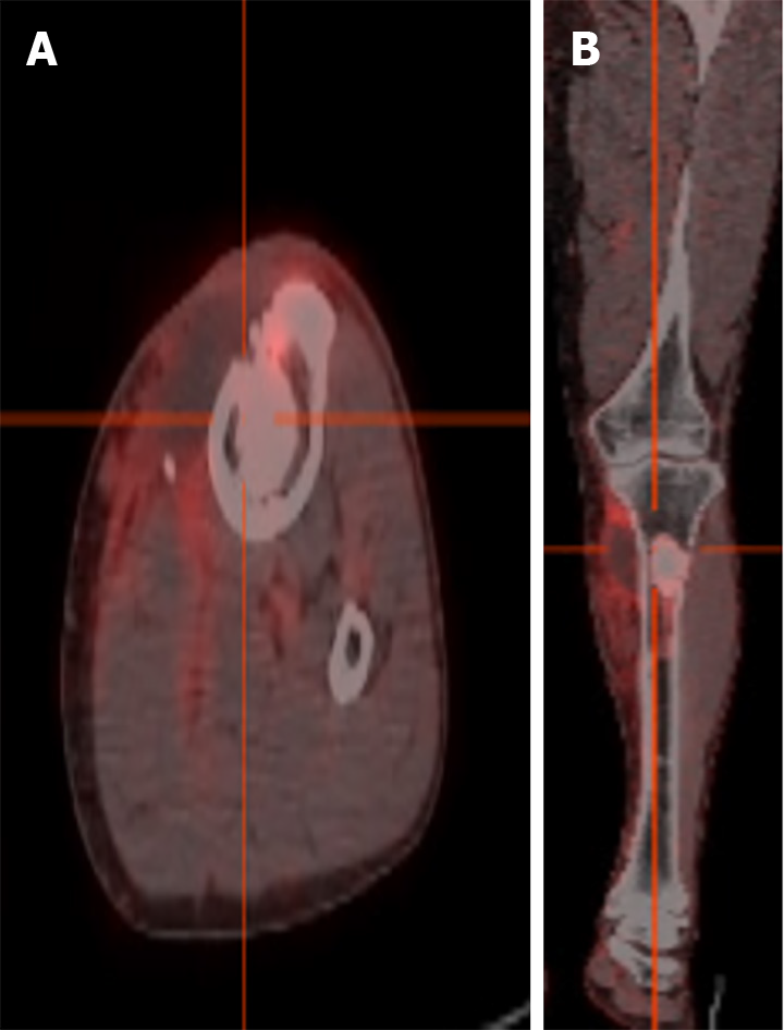
Figure 2 Positron emission tomography/computed tomography: Osteomyelitis of the upper tibia, local soft tissue infection.
A and B: A circular high-density shadow could be seen in the proximal tibia, with clear boundaries and locally increased fluorodeoxyglucose metabolism, shown in transverse (A) and coronal (B) views.
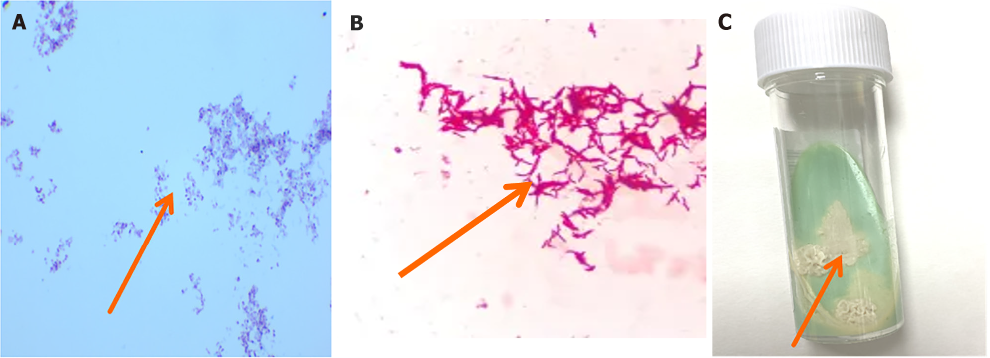
Figure 3 Cultivation of nontuberculous mycobacteria and microscopic images.
A: Positive acid fast staining of pus, with a large number of red elongated hyphae (100 ×); B: Under the microscope, slender, red, rod-shaped nontuberculous mycobacteria could be seen, with relatively scattered distribution and some bacterial cells slightly curved (400 ×); C: On the 12th day of observation, a unique scene appeared on Roche medium. Rice yellowish dry bacterial colonies appeared on top. This rice yellow color was very eye-catching and stood out, particularly against the background of the culture medium. The colony appeared dry and lacked a moist luster.
- Citation: Lin HY, Tan QH. Metagenomic next-generation sequencing may assist diagnosis of osteomyelitis caused by Mycobacterium houstonense: A case report. World J Orthop 2024; 15(11): 1095-1100
- URL: https://www.wjgnet.com/2218-5836/full/v15/i11/1095.htm
- DOI: https://dx.doi.org/10.5312/wjo.v15.i11.1095


