Copyright
©The Author(s) 2023.
World J Orthop. Feb 18, 2023; 14(2): 64-82
Published online Feb 18, 2023. doi: 10.5312/wjo.v14.i2.64
Published online Feb 18, 2023. doi: 10.5312/wjo.v14.i2.64
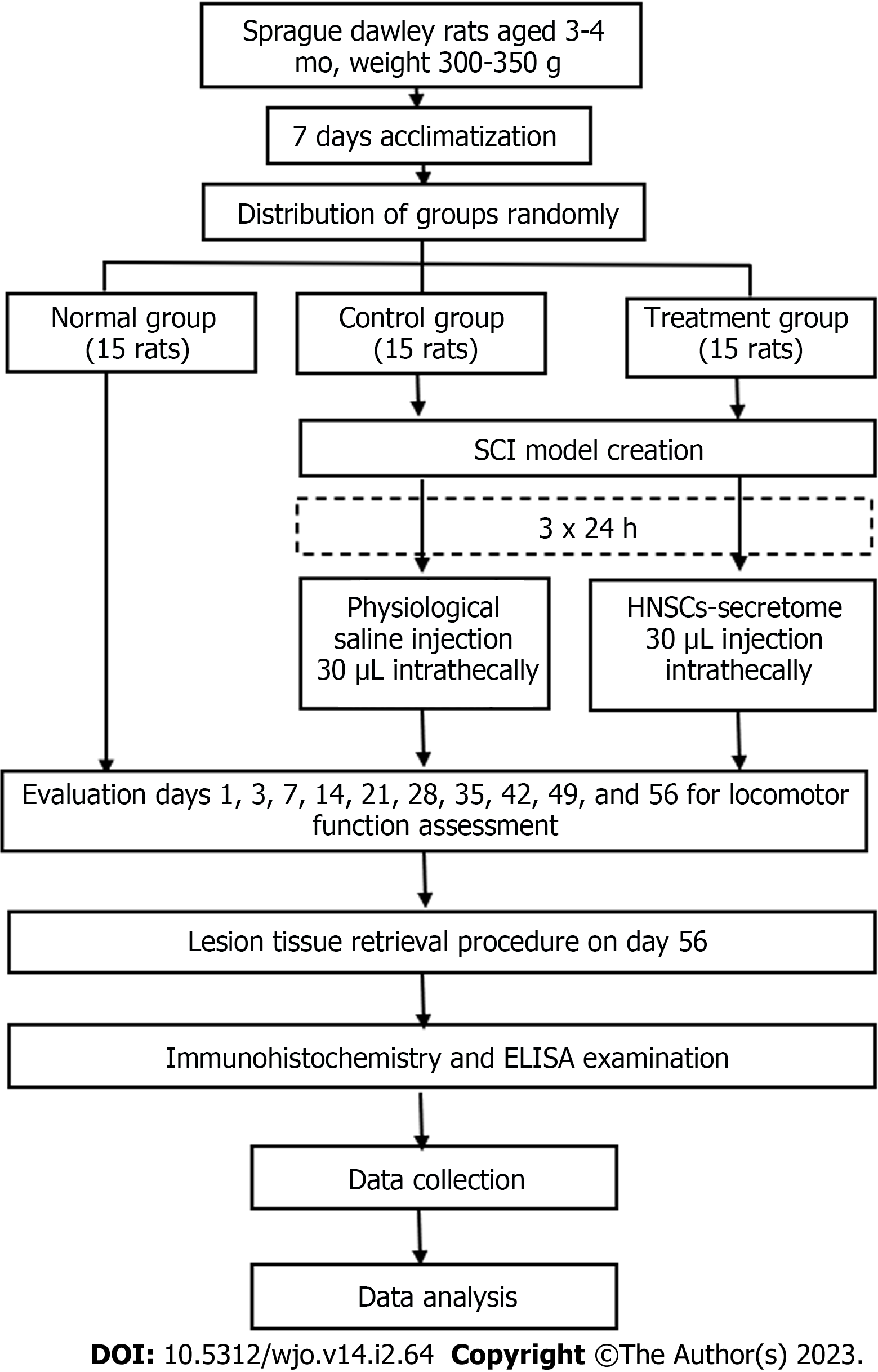
Figure 1 Diagram of the animal model and grouping.
SCI: Spinal cord injury; HNSCs: Human neural stem cells; ELISA: Enzyme-linked immunosorbent assay.
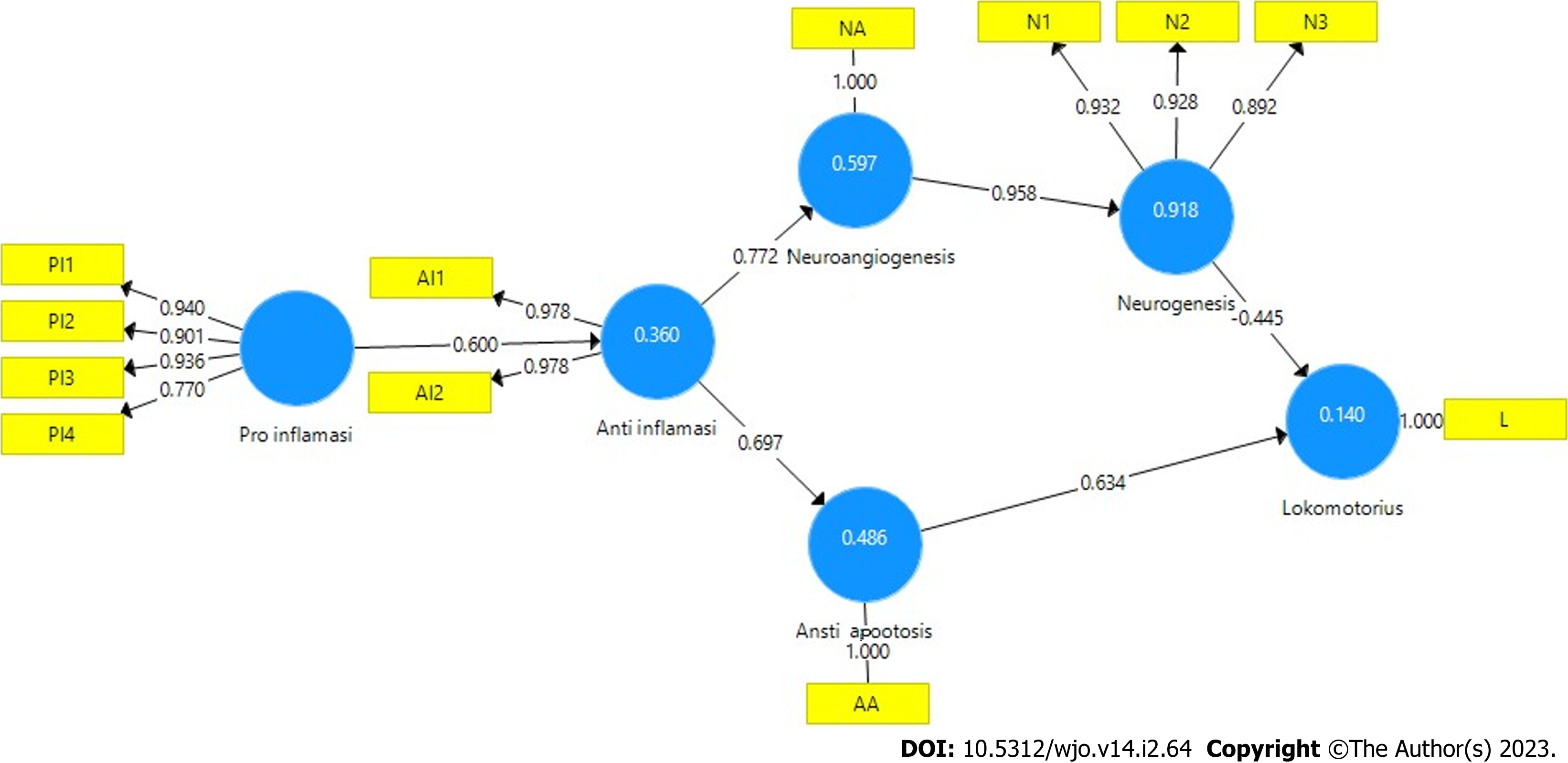
Figure 2 Diagram outer model based on partial least squares algorithm.
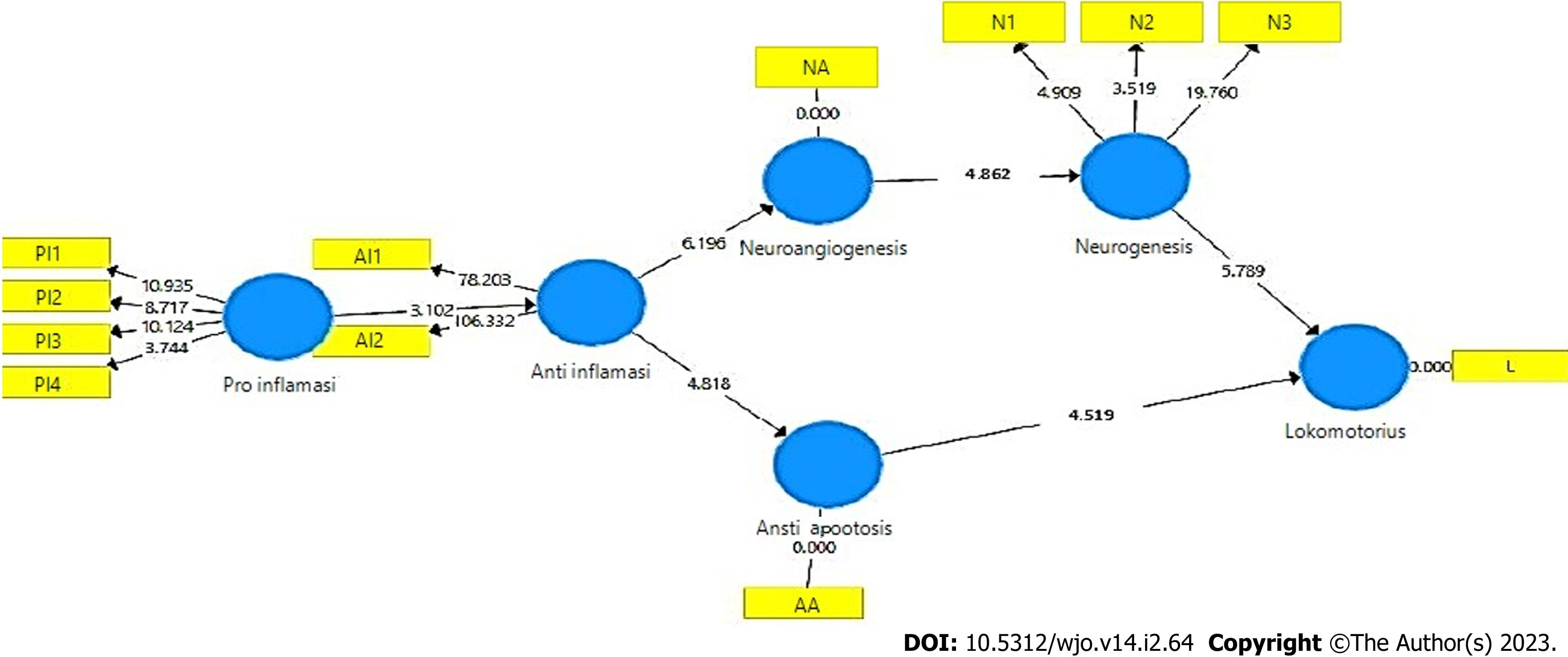
Figure 3 Diagram inner model using bootstrapping and blindfolding partial least squares structural equation modeling.
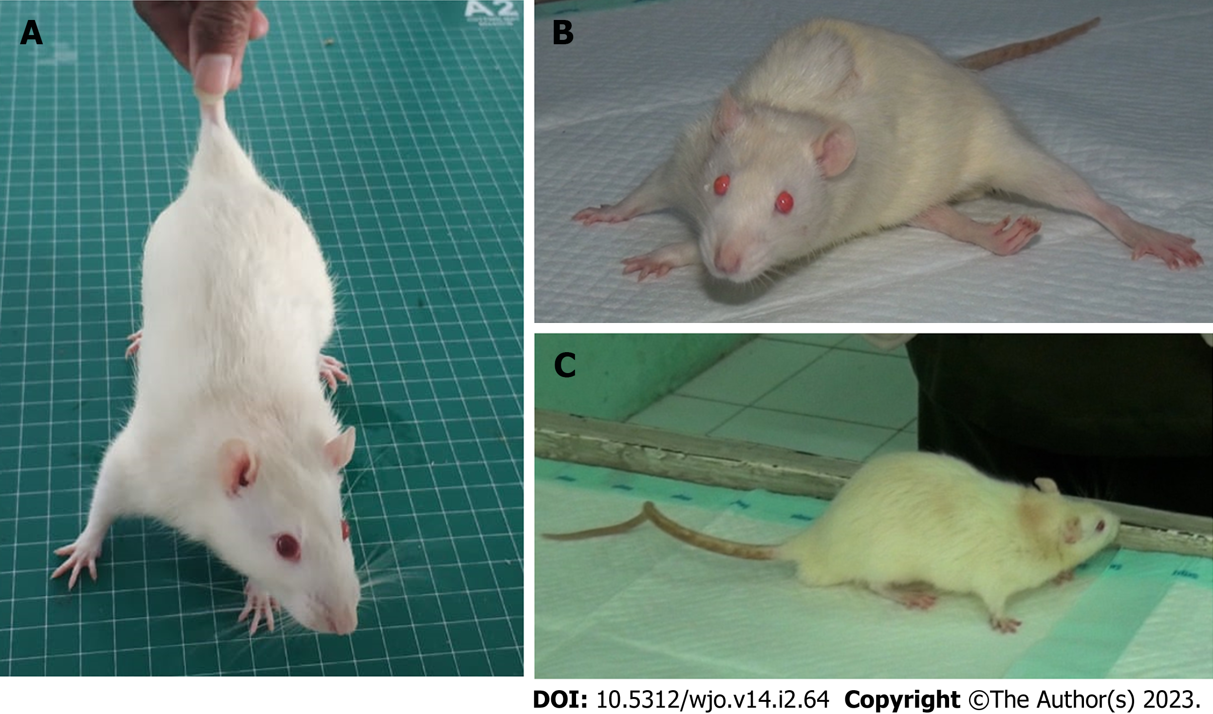
Figure 4 Evaluation Basso, Beattie, Bresnahan scores in different rat groups.
A: Normal group; B: Control group; C: Treatment group.
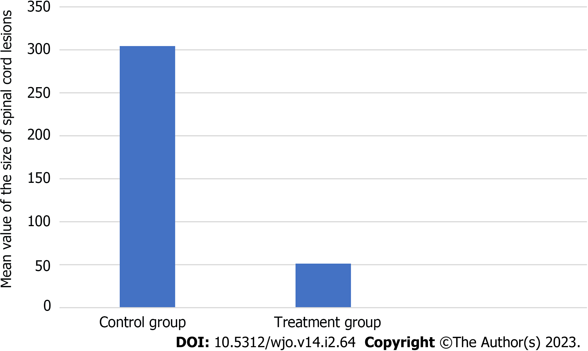
Figure 5 The mean value of the size of spinal cord lesions in the control and treatment groups.
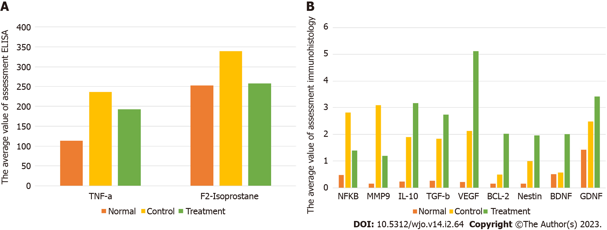
Figure 6 The mean value of biomarker by enzyme-linked immunosorbent assay and immuhistochemichal assessment.
A: Diagram showing the average value of enzyme-linked immunosorbent assay assessment; B: Diagram showing the average value of immuhistochemichal assessment. NF-kB: Nuclear factor-kappa B; MMP9: Metalloproteinase matrix-9; TNF-α: Tumor necrosis factor-α; IL-10: Interleukin-10; TGF-β: Transforming growth factor-β; VEGF: Vascular endothelial growth factor; Bcl-2: B-cell lymphoma 2; BDNF: Brain-derived neurotrophic factor; GDNF: Glial cell line-derived neurotrophic factor; F2-Isoprostanes: Free radical oxidative stress; ELISA: Enzyme-linked immunosorbent assay.
Figure 7 We observed immunohistochemical matrix metalloproteinase 9 average value of 10 field of views, every field of view have 625 µ2 with 400 × magnification.
A: Treatment group; B: Control group; C: Normal group. Microglia (red arrow) are small round cells, solid nuclei and give a positive reaction with anti matrix metalloproteinase 9 (MMP9) indicated by brown color. While macrophage (blue arrow) cells are large, vesicular nucleus, and sometimes elongated resembling fibroblasts (macrophages like fibroblast) and give a positive reaction with anti MMP9 indicated by brown.
Figure 8 We observed immunohistochemical transforming growth factor-β average value of 10 field of views, every field of view have 625 µ2 with 400× magnification.
A: Treatment group; B: Control group; C: Normal group. Microglia are small round cells, solid nuclei and give a positive reaction with anti-transforming growth factor-beta (TGF-β) indicated by brown color. While macrophage cells are large, vesicular nucleus,and sometimes elongated resembling fibroblasts (macrophages like fibroblast) and give a positive reaction with anti TGF-β indicated by brown.
Figure 9 We observed immunohistochemical vascular endothelial growth factor average value of 10 field of views, every field of view have 625 µ2 with 400 × magnification.
A: Treatment group; B: Control group; C: Normal group. Microglia are small round cells, solid nuclei and give a positive reaction with anti-vascular endothelial growth factor (VEGF) indicated by brown color. While macrophage cells are large, vesicular nucleus, and sometimes elongated resembling fibroblasts (macrophages like fibroblast) and give a positive reaction with anti-VEGF indicated by brown.
Figure 10 We observed immunohistochemical B cell lymphoma-2 average value of 10 field of views, every field of view have 625 µ2 with 400 × magnification.
A: Treatment group; B: Control group; C: Normal group. Microglia are small round cells, solid nuclei and give a positive reaction with anti B-cell lymphoma 2 (Bcl-2) indicated by brown color. While macrophage cells are large, vesicular nucleus, and sometimes elongated resembling fibroblasts (macrophages like fibroblast) and give a positive reaction with anti Bcl-2 indicated by brown.
Figure 11 We observed immunohistochemical brain derived neurotrophic factor average value of 10 field of views, every field of view have 625 µ2 with 400 × magnification.
A: Treatment group; B: Control group; C: Normal group. Normal group. Microglia are small round cells, solid nuclei and give a positive reaction with anti-brain derived neurotrophic factor (BDNF) indicated by brown color. While macrophage cells are large, vesicular nucleus, and sometimes elongated resembling fibroblasts (macrophages like fibroblast) and give a positive reaction with anti BDNF indicated by brown.
- Citation: Semita IN, Utomo DN, Suroto H. Mechanism of spinal cord injury regeneration and the effect of human neural stem cells-secretome treatment in rat model. World J Orthop 2023; 14(2): 64-82
- URL: https://www.wjgnet.com/2218-5836/full/v14/i2/64.htm
- DOI: https://dx.doi.org/10.5312/wjo.v14.i2.64


