Copyright
©The Author(s) 2022.
World J Orthop. Mar 18, 2022; 13(3): 278-288
Published online Mar 18, 2022. doi: 10.5312/wjo.v13.i3.278
Published online Mar 18, 2022. doi: 10.5312/wjo.v13.i3.278
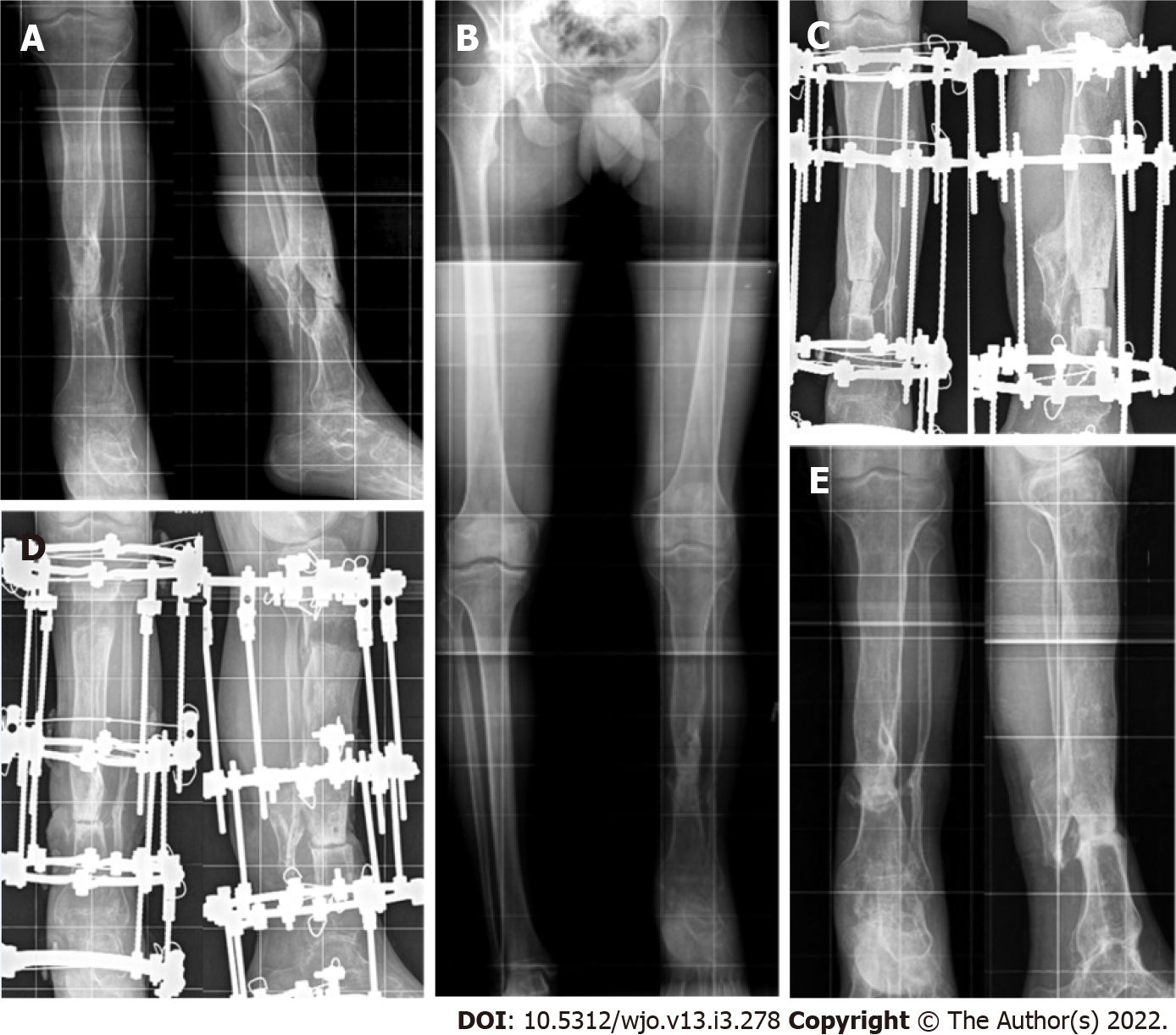
Figure 1 Post-traumatic defect case 4 (Table 1).
A: Preoperative radiographs of the right tibia capturing the adjacent joints showing a hypotrophic nonunion of the tibia; B: Preoperative telemetry compensated by a sole elevation 6-cm left leg discrepancy; C: Spacer fills the defect; D: Closed docking of the fragments and the regenerate of satisfactory optical density and zonal structure; E: Bone callus at the fragments docking and the regenerate with signs of its remodeling and cortical plates at 6-mo follow-up.
Figure 2 Congenital pseudarthrosis of the tibia case 3 (Table 2).
A: Preoperative radiographs of the left tibia capturing the adjacent joints showing valgus and antecurvatum at the pseudarthrosis level, extended sclerosis of fragments ends; B: Completion of distraction and defect filling at the time of docking between the ends without signs of ossification; C: Continuous distraction regenerate and consistent bone callus at the docking site at 1-year follow-up.
- Citation: Borzunov DY, Kolchin SN, Mokhovikov DS, Malkova TA. Ilizarov bone transport combined with the Masquelet technique for bone defects of various etiologies (preliminary results). World J Orthop 2022; 13(3): 278-288
- URL: https://www.wjgnet.com/2218-5836/full/v13/i3/278.htm
- DOI: https://dx.doi.org/10.5312/wjo.v13.i3.278


